ARG64143
anti-OGT antibody
anti-OGT antibody for Flow cytometry,ICC/IF,IHC-Formalin-fixed paraffin-embedded sections,Western blot and Human,Rat
Neuroscience antibody; Signaling Transduction antibody
Overview
| Product Description | Goat Polyclonal antibody recognizes OGT |
|---|---|
| Tested Reactivity | Hu, Rat |
| Predict Reactivity | Ms, Cow, Dog |
| Tested Application | FACS, ICC/IF, IHC-P, WB |
| Specificity | This antibody is expected to recognise both reported isoforms (NP_858058.1 and NP_858059.1 |
| Host | Goat |
| Clonality | Polyclonal |
| Isotype | IgG |
| Target Name | OGT |
| Antigen Species | Human |
| Immunogen | C-YEHPKDLKLSDGR |
| Conjugation | Un-conjugated |
| Alternate Names | UDP-N-acetylglucosamine--peptide N-acetylglucosaminyltransferase 110 kDa subunit; O-GLCNAC; EC 2.4.1.255; OGT; HRNT1; HINCUT-1; O-linked N-acetylglucosamine transferase 110 kDa subunit; O-GlcNAc transferase subunit p110 |
Application Instructions
| Application Suggestion |
|
||||||||||
|---|---|---|---|---|---|---|---|---|---|---|---|
| Application Note | WB: Recommend incubate at RT for 1h. IHC-P: Antigen Retrieval: Steam tissue section in Citrate buffer (pH 6.0). * The dilutions indicate recommended starting dilutions and the optimal dilutions or concentrations should be determined by the scientist. |
Properties
| Form | Liquid |
|---|---|
| Purification | Purified from goat serum by antigen affinity chromatography. |
| Buffer | Tris saline (pH 7.3), 0.02% Sodium azide and 0.5% BSA. |
| Preservative | 0.02% Sodium azide |
| Stabilizer | 0.5% BSA |
| Concentration | 0.5 mg/ml |
| Storage Instruction | For continuous use, store undiluted antibody at 2-8°C for up to a week. For long-term storage, aliquot and store at -20°C or below. Storage in frost free freezers is not recommended. Avoid repeated freeze/thaw cycles. Suggest spin the vial prior to opening. The antibody solution should be gently mixed before use. |
| Note | For laboratory research only, not for drug, diagnostic or other use. |
Bioinformation
| Database Links |
Swiss-port # O15294 Human UDP-N-acetylglucosamine--peptide N-acetylglucosaminyltransferase 110 kDa s Swiss-port # P56558 Rat UDP-N-acetylglucosamine--peptide N-acetylglucosaminyltransferase 110 kDa sub |
|---|---|
| Background | This gene encodes a glycosyltransferase that catalyzes the addition of a single N-acetylglucosamine in O-glycosidic linkage to serine or threonine residues. Since both phosphorylation and glycosylation compete for similar serine or threonine residues, the two processes may compete for sites, or they may alter the substrate specificity of nearby sites by steric or electrostatic effects. The protein contains multiple tetratricopeptide repeats that are required for optimal recognition of substrates. Alternatively spliced transcript variants encoding distinct isoforms have been found for this gene. [provided by RefSeq, Oct 2009] |
| Research Area | Neuroscience antibody; Signaling Transduction antibody |
| Calculated MW | 117 kDa |
| PTM | Ubiquitinated, leading to its proteasomal degradation. Phosphorylation on Ser-3 or Ser-4 by GSK3-beta positively regulates its activity. |
Images (6) Click the Picture to Zoom In
-

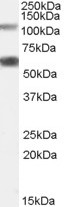
ARG64143 anti-OGT antibody WB image
Western Blot: Rat Pancreas lysate (35 µg protein in RIPA buffer) stained with ARG64143 anti-OGT antibody at 0.05 µg/ml dilution.
-
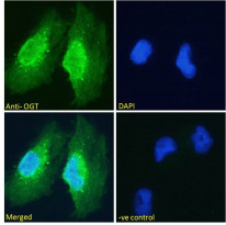
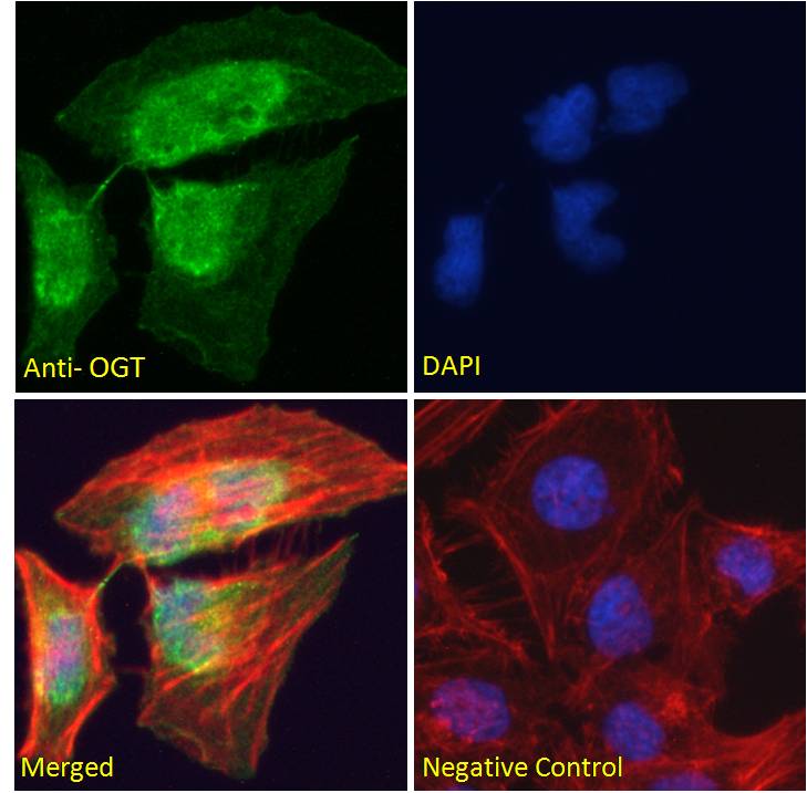
ARG64143 anti-OGT antibody ICC/IF image
Immunofluorescence: Paraformaldehyde fixed U251 cells permeabilized with 0.15% Triton. Cells were stained with ARG64143 anti-OGT antibody (green) at 10 µg/ml dilution for 1 hour. DAPI (blue) for nuclear staining. Negative control: Unimmunized goat IgG (green) at 10 µg/ml dilution.
-

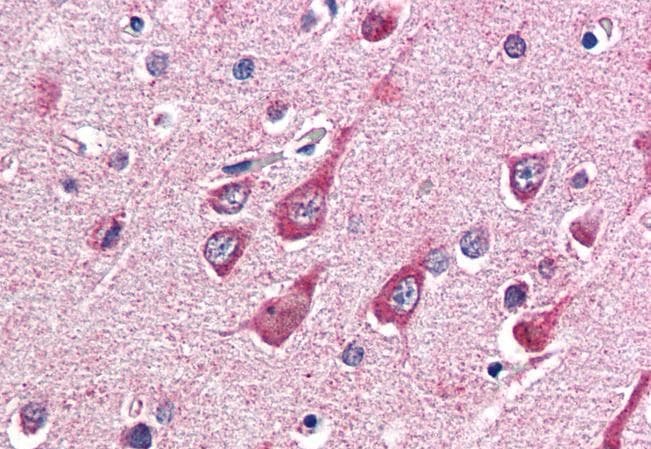
ARG64143 anti-OGT antibody IHC-P image
Immunohistochemistry: Paraffin-embedded Human cortex tissue. Antigen Retrieval: Steam tissue section in Citrate buffer (pH 6.0). The tissue section was stained with ARG64143 anti-OGT antibody at 5 µg/ml dilution followed by AP-staining.
-
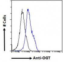
ARG64143 anti-OGT antibody FACS image
Flow Cytometry: Paraformaldehyde-fixed HEK293 cells permeabilized with 0.5% Triton. Cells were stained with ARG64143 anti-OGT antibody (blue line) at 10 µg/ml dilution for 1 hour, followed by incubation with Alexa FluorR 488 labelled secondary antibody. IgG control: Unimmunized goat IgG (black line).
-

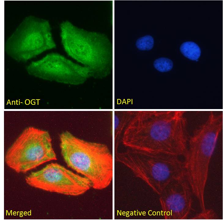
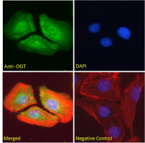
ARG64143 anti-OGT antibody ICC/IF image
Immunofluorescence: Paraformaldehyde fixed HeLa cells permeabilized with 0.15% Triton. Cells were stained with ARG64143 anti-OGT antibody (green) at 10 µg/ml dilution for 1 hour. DAPI (blue) for nuclear staining. Phalloidin (red) for Actin filaments staining. Negative control: Unimmunized goat IgG (green) at 10 µg/ml dilution.
-
ARG64143 anti-OGT antibody ICC/IF image
Immunofluorescence: Paraformaldehyde fixed U2OS cells permeabilized with 0.15% Triton. Cells were stained with ARG64143 anti-OGT antibody (green) at 10 µg/ml dilution for 1 hour. DAPI (blue) for nuclear staining. Phalloidin (red) for Actin filaments staining. Negative control: Unimmunized goat IgG (green) at 10 µg/ml dilution.