ARG66559
anti-Neurotensin Receptor 1 antibody
anti-Neurotensin Receptor 1 antibody for ICC/IF,IHC-Formalin-fixed paraffin-embedded sections,Western blot and Human
Overview
| Product Description | Rabbit Polyclonal antibody recognizes Neurotensin Receptor 1 |
|---|---|
| Tested Reactivity | Hu |
| Tested Application | ICC/IF, IHC-P, WB |
| Host | Rabbit |
| Clonality | Polyclonal |
| Isotype | IgG |
| Target Name | Neurotensin Receptor 1 |
| Antigen Species | Human |
| Immunogen | KLH-conjugated synthetic peptide encompassing a sequence within the center region of Human NTR1. |
| Conjugation | Un-conjugated |
| Alternate Names | NTR; NTR1; Neurotensin receptor type 1; NT-R-1; NTRH; High-affinity levocabastine-insensitive neurotensin receptor |
Application Instructions
| Application Suggestion |
|
||||||||
|---|---|---|---|---|---|---|---|---|---|
| Application Note | IHC-P: Antigen Retrieval: Heat mediation was performed in Sodium citrate buffer (pH 6.0). * The dilutions indicate recommended starting dilutions and the optimal dilutions or concentrations should be determined by the scientist. |
||||||||
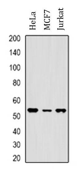
| Observed Size | 54 kDa |
Properties
| Form | Liquid |
|---|---|
| Purification | Affinity purification with immunogen. |
| Buffer | 0.42% Potassium phosphate (pH 7.3), 0.87% NaCl, 0.01% Sodium azide and 30% Glycerol. |
| Preservative | 0.01% Sodium azide |
| Stabilizer | 30% Glycerol |
| Storage Instruction | For continuous use, store undiluted antibody at 2-8°C for up to a week. For long-term storage, aliquot and store at -20°C. Storage in frost free freezers is not recommended. Avoid repeated freeze/thaw cycles. Suggest spin the vial prior to opening. The antibody solution should be gently mixed before use. |
| Note | For laboratory research only, not for drug, diagnostic or other use. |
Bioinformation
| Database Links | |
|---|---|
| Gene Symbol | NTSR1 |
| Gene Full Name | neurotensin receptor 1 (high affinity) |
| Background | Neurotensin receptor 1 belongs to the large superfamily of G-protein coupled receptors. NTSR1 mediates the multiple functions of neurotensin, such as hypotension, hyperglycemia, hypothermia, antinociception, and regulation of intestinal motility and secretion. [provided by RefSeq, Jul 2008] |
| Function | G-protein coupled receptor for the tridecapeptide neurotensin (NTS). Signaling is effected via G proteins that activate a phosphatidylinositol-calcium second messenger system. Signaling leads to the activation of downstream MAP kinases and protects cells against apoptosis. [UniProt] |
| Cellular Localization | Cell membrane; Multi-pass membrane protein. Membrane raft. Note=Palmitoylation is required for localization at CAV1-enriched membrane rafts. [UniProt] |
| Calculated MW | 46 kDa |
| PTM | N-glycosylated. Palmitoylated; this is required for normal localization at membrane rafts and normal GNA11-mediated activation of down-stream signaling cascades. The palmitoylation level increases in response to neurotensin treatment. [UniProt] |
Images (3) Click the Picture to Zoom In
-
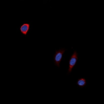
ARG66559 anti-Neurotensin Receptor 1 antibody ICC/IF image
Immunofluorescence: Formalin-fixed SK-N-SH cells were permeabilized with 0.1% Triton X-100 in TBS for 5-10 minutes and blocked with 3% BSA-PBS for 30 minutes at room temperature. Cells were stained with ARG66559 anti-Neurotensin Receptor 1 antibody (red) in 3% BSA-PBS and incubated overnight at 4°C in a hidified chamber. DAPI was used to stain the cell nuclei (blue).
-
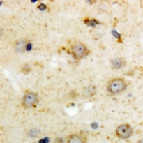
ARG66559 anti-Neurotensin Receptor 1 antibody IHC-P image
Immunohistochemistry: Formalin-fixed and paraffin-embedded Human brain tissue. Antigen Retrieval: Heat mediation was performed in Sodium citrate buffer (pH 6.0). The section was then stained with ARG66559 anti-Neurotensin Receptor 1 antibody at room temperature and detected using an HRP conjugacompact polymer system. DAB was used as the chromogen. The section was then counterstained with haematoxylin and mounted with DPX.
-

ARG66559 anti-Neurotensin Receptor 1 antibody WB image
Western blot: HeLa, MCF7 and Jurkat whole cell lysates stained with ARG66559 anti-Neurotensin Receptor 1 antibody.