ARG63076
anti-Neurofilament NF-M antibody [NF-09]
anti-Neurofilament NF-M antibody [NF-09] for ICC/IF,IHC-Formalin-fixed paraffin-embedded sections,Western blot and Human,Mouse,Rat,Mammal
Controls and Markers antibody; Developmental Biology antibody; Neuroscience antibody; Signaling Transduction antibody; Intermediate Neurofilament antibody
Overview
| Product Description | Mouse Monoclonal antibody [NF-09] recognizes Neurofilament NF-M |
|---|---|
| Tested Reactivity | Hu, Ms, Rat, Mamm |
| Tested Application | ICC/IF, IHC-P, WB |
| Specificity | The clone NF-09 reacts with both phosphorylated and non-phosphorylated form of medium neurofilament protein (160 kDa) of various species. |
| Host | Mouse |
| Clonality | Monoclonal |
| Clone | NF-09 |
| Isotype | IgG2a |
| Target Name | Neurofilament NF-M |
| Antigen Species | Pig |
| Immunogen | Pellet of porcine brain cold stable proteins after depolymerization of microtubules. |
| Conjugation | Un-conjugated |
| Alternate Names | Neurofilament medium polypeptide; Neurofilament 3; Neurofilament triplet M protein; NFM; NF-M; 160 kDa neurofilament protein; NEF3 |
Application Instructions
| Application Suggestion |
|
||||||||
|---|---|---|---|---|---|---|---|---|---|
| Application Note | ICC/IF: Carnoys fixative 2 x 3 min, blocking 1% glycine + 0.2% gelatin 10 min. * The dilutions indicate recommended starting dilutions and the optimal dilutions or concentrations should be determined by the scientist. |
||||||||
| Positive Control | ICC/IF: Neuro2A |
Properties
| Form | Liquid |
|---|---|
| Purification | Purified from ascites by protein-A affinity chromatography. |
| Purity | > 95% (by SDS-PAGE) |
| Buffer | PBS (pH 7.4) and 15 mM Sodium azide |
| Preservative | 15 mM Sodium azide |
| Concentration | 1 mg/ml |
| Storage Instruction | For continuous use, store undiluted antibody at 2-8°C for up to a week. For long-term storage, aliquot and store at -20°C or below. Storage in frost free freezers is not recommended. Avoid repeated freeze/thaw cycles. Suggest spin the vial prior to opening. The antibody solution should be gently mixed before use. |
| Note | For laboratory research only, not for drug, diagnostic or other use. |
Bioinformation
| Database Links | |
|---|---|
| Background | Neurofilaments (NFs) are a type of intermediate filament (IF) expressed almost exclusively in neuronal cells, and in those cells most prominently in large axons. NFs in most vertebrates are composed of three different polypeptide chains with different molecular weights – neurofilament medium protein (NF-M), high (NF-H) and light protein (NF-L), which share sequence and structural similarity in a coiled-coil core domain, but differ in the length and sequence of their N-termini and more dramatically of their C-termini which in the case of NF-M and NF-H form the flexible extensions that link NFs to each other and to other elements in the cytoplasm. NF-M protein tail-mediated interactions of neurofilaments are critical for size and cytoskeletal architecture of axons, and are mediated, in part, by the highly phosphorylated tail domain of this protein. NF-M phosphorylation and O-GlcNAcylation are regulated reciprocally and affect its translocation and filament formation and function. Antibodies to the various neurofilament subunits are very useful cell type markers since the proteins are among the most abundant of the nervous system, are expressed only in neurons and are biochemically very stable. |
| Research Area | Controls and Markers antibody; Developmental Biology antibody; Neuroscience antibody; Signaling Transduction antibody; Intermediate Neurofilament antibody |
| Calculated MW | 102 kDa |
| PTM | There are a number of repeats of the tripeptide K-S-P, NFM is phosphorylated on a number of the serines in this motif. It is thought that phosphorylation of NFM results in the formation of interfilament cross bridges that are important in the maintenance of axonal caliber. Phosphorylation seems to play a major role in the functioning of the larger neurofilament polypeptides (NF-M and NF-H), the levels of phosphorylation being altered developmentally and coincidentally with a change in the neurofilament function. Phosphorylated in the head and rod regions by the PKC kinase PKN1, leading to the inhibition of polymerization. |
Images (2) Click the Picture to Zoom In
-
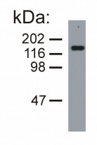
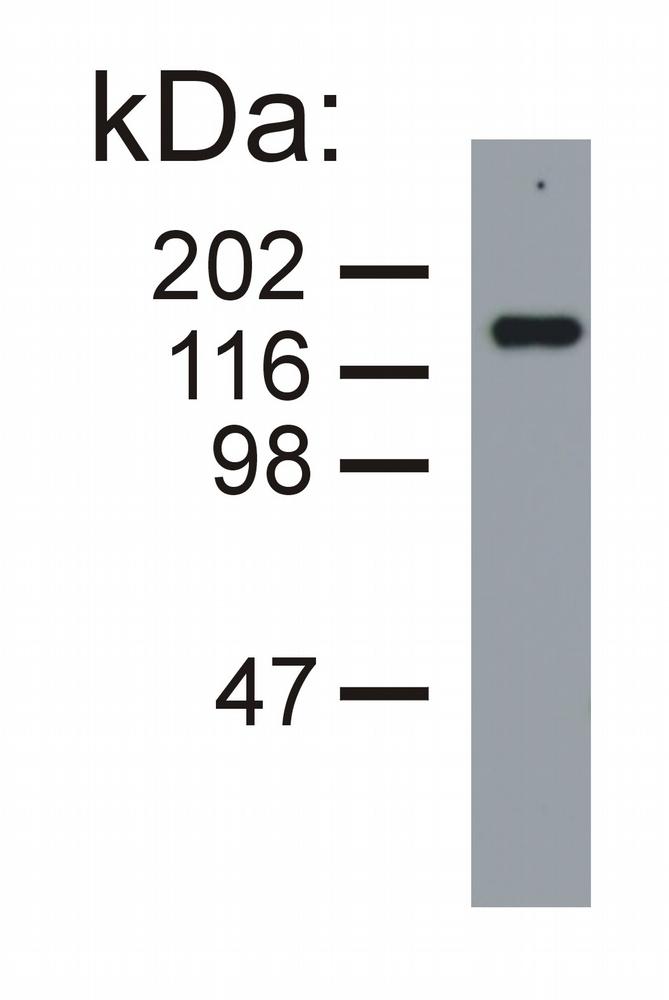
ARG63076 anti-Neurofilament NF-M antibody [NF-09] WB image
Western blot: Porcine brain lysate (under reducing conditions) stained with ARG63076 anti-Neurofilament NF-M antibody [NF-09].
-
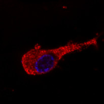
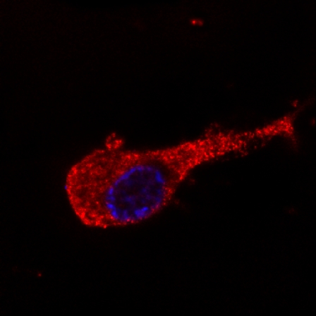
ARG63076 anti-Neurofilament NF-M antibody [NF-09] ICC/IF image
Immunofluorescence: Murine Neuro2A cells stained with ARG63076 anti-Neurofilament NF-M antibody [NF-09] (green).
Cell nuclei was stained with DAPI (blue).