ARG10724
anti-Neurofilament NF-M antibody
anti-Neurofilament NF-M antibody for ICC/IF,IHC-Frozen sections,Western blot and Human,Mouse,Rat,Cow,Horse,Pig
Controls and Markers antibody; Developmental Biology antibody; Neuroscience antibody; Signaling Transduction antibody; Intermediate Neurofilament antibody
Overview
| Product Description | Rabbit Polyclonal antibody recognizes Neurofilament NF-M |
|---|---|
| Tested Reactivity | Hu, Ms, Rat, Cow, Hrs, Pig |
| Predict Reactivity | Chk |
| Tested Application | ICC/IF, IHC-Fr, WB |
| Host | Rabbit |
| Clonality | Polyclonal |
| Isotype | IgG |
| Target Name | Neurofilament NF-M |
| Antigen Species | Rat |
| Immunogen | Recombinant fusion protein containing the extreme C-terminal segment of Rat NF-M. |
| Conjugation | Un-conjugated |
| Alternate Names | Neurofilament medium polypeptide; Neurofilament 3; Neurofilament triplet M protein; NFM; NF-M; 160 kDa neurofilament protein; NEF3 |
Application Instructions
| Application Suggestion |
|
||||||||
|---|---|---|---|---|---|---|---|---|---|
| Application Note | * The dilutions indicate recommended starting dilutions and the optimal dilutions or concentrations should be determined by the scientist. |
Properties
| Form | Liquid |
|---|---|
| Purification | Unpurified. |
| Buffer | Serum. |
| Storage Instruction | For continuous use, store undiluted antibody at 2-8°C for up to a week. For long-term storage, aliquot and store at -20°C or below. Storage in frost free freezers is not recommended. Avoid repeated freeze/thaw cycles. Suggest spin the vial prior to opening. The antibody solution should be gently mixed before use. |
| Note | For laboratory research only, not for drug, diagnostic or other use. |
Bioinformation
| Database Links | |
|---|---|
| Gene Symbol | Nefm |
| Gene Full Name | neurofilament, medium polypeptide |
| Background | Neurofilaments are type IV intermediate filament heteropolymers composed of light, medium, and heavy chains. Neurofilaments comprise the axoskeleton and functionally maintain neuronal caliber. They may also play a role in intracellular transport to axons and dendrites. This gene encodes the medium neurofilament protein. This protein is commonly used as a biomarker of neuronal damage. Alternative splicing results in multiple transcript variants encoding distinct isoforms. [provided by RefSeq, Oct 2008] |
| Function | Neurofilaments usually contain three intermediate filament proteins: L, M, and H which are involved in the maintenance of neuronal caliber. [UniProt] |
| Research Area | Controls and Markers antibody; Developmental Biology antibody; Neuroscience antibody; Signaling Transduction antibody; Intermediate Neurofilament antibody |
| Calculated MW | 102 kDa |
| PTM | There are a number of repeats of the tripeptide K-S-P, NFM is phosphorylated on a number of the serines in this motif. It is thought that phosphorylation of NFM results in the formation of interfilament cross bridges that are important in the maintenance of axonal caliber. Phosphorylation seems to play a major role in the functioning of the larger neurofilament polypeptides (NF-M and NF-H), the levels of phosphorylation being altered developmentally and coincidentally with a change in the neurofilament function. Phosphorylated in the head and rod regions by the PKC kinase PKN1, leading to the inhibition of polymerization. |
Images (4) Click the Picture to Zoom In
-
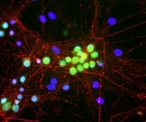
ARG10724 anti-Neurofilament NF-M antibody ICC/IF image
Immunocytochemistry: Rat mixed neuron / glia cultures stained with ARG10724 anti-Neurofilament NF-M antibody (red) and co-stained with Mouse monoclonal antibody [1G10] to Fox1 (green). ARG10724 anti-Neurofilament NF-M antibody stains axonal, dendritic and perikaryal profiles of neurons cleanly and specifically. Like antibody to Fox3 / NeuN, the Fox1 antibody binds to the nuclei of neurons only. DNA is shown in blue with DAPI.
-

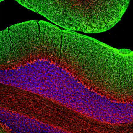
ARG10724 anti-Neurofilament NF-M antibody IHC-Fr image
Immunohistochemistry: Frozen section of Rat cerebellum stained with ARG10724 anti-Neurofilament NF-M antibody (red) at 1:2000 dilution and costained with ARG10715 anti-GAP43 antibody [3H14] (green) at 1:2000 dilution. (Sample preparation: Following transcardial perfusion of Rat with 4% paraformaldehyde, brain was post fixed for 24 hours, cut to 45 µM, and free-floating sections were stained with the above antibodies.)
The NF-M antibody strongly labels neuronal processes throughout the cerebellum, while the GAP43 antibody stains predominantly synaptic regions in the molecular layer.
-
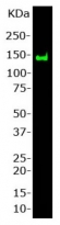
ARG10724 anti-Neurofilament NF-M antibody WB image
Western blot: Rat spinal cord stained with ARG10724 anti-Neurofilament NF-M antibody at 1:1000 dilution. A prominent band running with an apparent SDS-PAGE molecular weight of ~145 kDa corresponds to rodent NF-M.
-
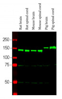
ARG10724 anti-Neurofilament NF-M antibody WB image
Western blot: Rat brain, Rat spinal cord, Mouse brain, Mouse spinal cord, Pig brain and Pig spinal cord lysates stained with ARG10724 anti-Neurofilament NF-M antibody (green) at 1:2000 dilution.