ARG52350
anti-Neurofilament NF-M antibody [3H11]
anti-Neurofilament NF-M antibody [3H11] for ICC/IF,IHC-Frozen sections,Western blot and Cow,Mouse,Rat
Controls and Markers antibody; Developmental Biology antibody; Neuroscience antibody; Signaling Transduction antibody; Intermediate Neurofilament antibody
Overview
| Product Description | Mouse Monoclonal antibody [3H11] recognizes Neurofilament NF-M |
|---|---|
| Tested Reactivity | Ms, Rat, Cow |
| Predict Reactivity | Hu, Chk |
| Tested Application | ICC/IF, IHC-Fr, WB |
| Host | Mouse |
| Clonality | Monoclonal |
| Clone | 3H11 |
| Isotype | IgG1 |
| Target Name | Neurofilament NF-M |
| Antigen Species | Rat |
| Immunogen | Preparation containing the extreme C-terminus expressed in and purified from E. Coli |
| Conjugation | Un-conjugated |
| Alternate Names | Neurofilament medium polypeptide; Neurofilament 3; Neurofilament triplet M protein; NFM; NF-M; 160 kDa neurofilament protein; NEF3 |
Application Instructions
| Application Suggestion |
|
||||||||
|---|---|---|---|---|---|---|---|---|---|
| Application Note | Specific for the ~145k neurofilament M protein. * The dilutions indicate recommended starting dilutions and the optimal dilutions or concentrations should be determined by the scientist. |
Properties
| Form | Liquid |
|---|---|
| Purification | Total IgG fraction |
| Buffer | Total IgG fraction and 10 mM Sodium azide |
| Preservative | 10 mM Sodium azide |
| Storage Instruction | For continuous use, store undiluted antibody at 2-8°C for up to a week. For long-term storage, aliquot and store at -20°C or below. Storage in frost free freezers is not recommended. Avoid repeated freeze/thaw cycles. Suggest spin the vial prior to opening. The antibody solution should be gently mixed before use. |
| Note | For laboratory research only, not for drug, diagnostic or other use. |
Bioinformation
| Database Links | |
|---|---|
| Gene Symbol | NEFM |
| Gene Full Name | neurofilament, medium polypeptide |
| Background | Neurofilaments are the 10nm or intermediate filament proteins found specifically in neurons, and are composed predominantly of three major proteins called NF-L, NF-M and NF-H . NF-M is the neurofilament middle or medium molecular weight polypeptide and runs on SDS-PAGE gels at 145-160 kDa, with some variability across species boundaries. Antibodies to NF-M are useful for identifying neuronal cells and their processes in tissue sections and in tissue culture. NF-M antibodies can also be useful to visualize neurofilament accumulations seen in many neurological diseases, such as Amyotrophic Lateral Sclerosis (Lou Gehrig's disease) and Alzheimer's disease . |
| Highlight | Related products: Neurofilament NF M antibodies; Neurofilament NF M Duos / Panels; Anti-Mouse IgG secondary antibodies; Related news: Neuronal Development Marker |
| Research Area | Controls and Markers antibody; Developmental Biology antibody; Neuroscience antibody; Signaling Transduction antibody; Intermediate Neurofilament antibody |
| Calculated MW | 102 kDa |
| PTM | There are a number of repeats of the tripeptide K-S-P, NFM is phosphorylated on a number of the serines in this motif. It is thought that phosphorylation of NFM results in the formation of interfilament cross bridges that are important in the maintenance of axonal caliber. Phosphorylation seems to play a major role in the functioning of the larger neurofilament polypeptides (NF-M and NF-H), the levels of phosphorylation being altered developmentally and coincidentally with a change in the neurofilament function. Phosphorylated in the head and rod regions by the PKC kinase PKN1, leading to the inhibition of polymerization. |
Images (4) Click the Picture to Zoom In
-
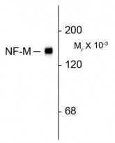
ARG52350 anti-Neurofilament NF-M antibody [3H11] WB image
Western Blot: rat cortex lysate showing specific immunolabeling of the ~145k NF-M protein stained with ARG52350 anti-Neurofilament NF-M antibody [3H11]
-
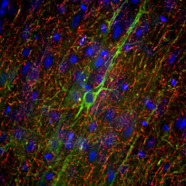
ARG52350 anti-Neurofilament NF-M antibody [3H11] IHC-Fr image
Immunohistochemistry: Frozen section of adult Rat frontal cortex tissue stained with ARG52350 anti-Neurofilament NF-M antibody [3H11] (green) at 1:5000 dilution, and costained with ARG52347 anti-Neurofilament NF-H antibody (red) at 1:5000 dilution. Following transcardial perfusion of Rat with 4% paraformaldehyde, brain was post fixed for 24 hours, cut to 45 µM, and free-floating sections were stained with above antibodies.
Clone 3H11 labels neuron cell bodies and dendrites of pyramidal neurons, as well as dendrites and axons of other neuronal cells, while the NF-H antibody stains the network of neuronal axons only.
-
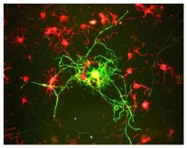
ARG52350 anti-Neurofilament NF-M antibody [3H11] ICC/IF image
Immunofluorescence: cultured rat neurons stained with ARG52350 anti-Neurofilament NF-M antibody [3H11] showing labeling of NF-M (green) in mature neurons.
-
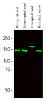
ARG52350 anti-Neurofilament NF-M antibody [3H11] WB image
Western blot: Rat spinal cord, Mouse spinal cord, Cow spinal cord and Rat sciatic nerve lysates stained with ARG52350 anti-Neurofilament NF-M antibody [3H11] (green) at 1:10000 dilution.


Clone References
Gamma-diketone axonopathy: analyses of cytoskeletal motors and highways in CNS myelinated axons.
WB / Rat


Enhanced cisplatin cytotoxicity by disturbing the nucleotide excision repair pathway in ovarian cancer cell lines.
WB / Human
Preferential transformation of human neuronal cells by human adenoviruses and the origin of HEK 293 cells.
ICC/IF / Human
Compartmentation of alpha-internexin and neurofilament triplet proteins in cultured hippocampal neurons.
WB / Rat


