ARG52349
anti-Neurofilament NF-L antibody
anti-Neurofilament NF-L antibody for ICC/IF,IHC-Frozen sections,Western blot and Human,Mouse,Rat
Controls and Markers antibody; Developmental Biology antibody; Neuroscience antibody; Signaling Transduction antibody; Neuronal Cytoskeletal antibody; Neurofilament antibody; Intermediate Neurofilament antibody
Overview
| Product Description | Chicken Polyclonal antibody recognizes Neurofilament NF-L |
|---|---|
| Tested Reactivity | Hu, Ms, Rat |
| Predict Reactivity | Chk |
| Tested Application | ICC/IF, IHC-Fr, WB |
| Host | Chicken |
| Clonality | Polyclonal |
| Isotype | IgY |
| Target Name | Neurofilament NF-L |
| Antigen Species | Bovine |
| Immunogen | Preparation of bovine spinal cord NF-L |
| Conjugation | Un-conjugated |
| Alternate Names | Neurofilament triplet L protein; 68 kDa neurofilament protein; CMT1F; NF68; NFL; CMT2E; Neurofilament light polypeptide; NF-L; PPP1R110 |
Application Instructions
| Application Suggestion |
|
||||||||
|---|---|---|---|---|---|---|---|---|---|
| Application Note | Specific for the ~ 68 kDa Neurofilament L protein in Western blots and works well on frozen sections, cells in tissue culture and on mildly formalin fixed histological sections. * The dilutions indicate recommended starting dilutions and the optimal dilutions or concentrations should be determined by the scientist. |
||||||||
| Observed Size | ~ 68 kDa |
Properties
| Form | Liquid |
|---|---|
| Purification | Total IgY fraction |
| Buffer | Total IgY fraction in PBS and 10 mM Sodium azide |
| Preservative | 10 mM Sodium azide |
| Storage Instruction | For continuous use, store undiluted antibody at 2-8°C for up to a week. For long-term storage, aliquot and store at -20°C or below. Storage in frost free freezers is not recommended. Avoid repeated freeze/thaw cycles. Suggest spin the vial prior to opening. The antibody solution should be gently mixed before use. |
| Note | For laboratory research only, not for drug, diagnostic or other use. |
Bioinformation
| Database Links | |
|---|---|
| Gene Symbol | NEFL |
| Gene Full Name | neurofilament, light polypeptide |
| Background | Neurofilaments are the 10nm or intermediate filament proteins found specifically in neurons, and are composed predominantly of three major proteins called NF-L, NF-M and NF-H . NF-L is the neurofilament light or low molecular weight polypeptide and runs on SDS-PAGE gels at about 68kDa. Antibodies to NF-L are useful for identifying neuronal cells and their processes in tissue sections and in tissue culture. Mutations in the protein coding region of the human NF-L gene cause some forms of Charcot-Marie-Tooth disease . |
| Function | Neurofilaments usually contain three intermediate filament proteins: L, M, and H which are involved in the maintenance of neuronal caliber. [UniProt] |
| Research Area | Controls and Markers antibody; Developmental Biology antibody; Neuroscience antibody; Signaling Transduction antibody; Neuronal Cytoskeletal antibody; Neurofilament antibody; Intermediate Neurofilament antibody |
| Calculated MW | 62 kDa |
| PTM | O-glycosylated. Phosphorylated in the head and rod regions by the PKC kinase PKN1, leading to the inhibition of polymerization. Ubiquitinated in the presence of TRIM2 and UBE2D1. |
Images (4) Click the Picture to Zoom In
-
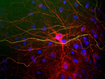
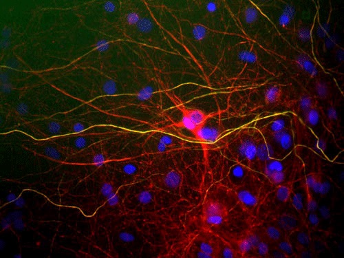
ARG52349 anti-Neurofilament NF-L antibody ICC/IF image
Immunofluorescence: Mixed cultured rat neurons and glia stained with ARG52349 anti-Neurofilament NF-L antibody (red) at 1:5000 dilution.
The NF-L protein is assembled into neurofilaments which are found throughout the axons, dendrites, and perikarya.
-
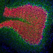
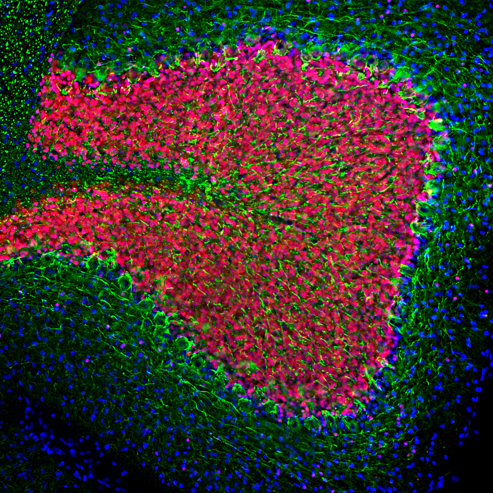
ARG52349 anti-Neurofilament NF-L antibody IHC-Fr image
Immunohistochemistry: Frozen section of Rat cerebellum tissue stained with ARG52349 anti-Neurofilament NF-L antibody (green) at 1:2000 dilution, and co-stained with ARG52283 anti-FOX3 / NeuN antibody [1B7] (red) at 1:5000 dilution. (Sample preparation: Following transcardial perfusion of Rat with 4% paraformaldehyde, brain was post fixed for 24 hours, cut to 45 µM, and free-floating sections were stained with the above antibodies.)
The NF-L antibody labels perikarya and processes of neuronal cells, particularly strongly the axons of basket cells, while the FOX3 / NeuN antibody stains the nuclei and proximal cytoplasm of neurons.
-
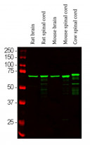
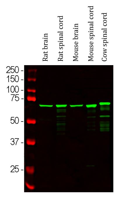
ARG52349 anti-Neurofilament NF-L antibody WB image
Western blot: Rat brain, Rat spinal cord, Mouse brain, Mouse spinal cord and Cow spinal cord lysates stained with ARG52349 anti-Neurofilament NF-L antibody (green) at 1:20000 dilution.
-
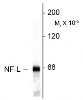
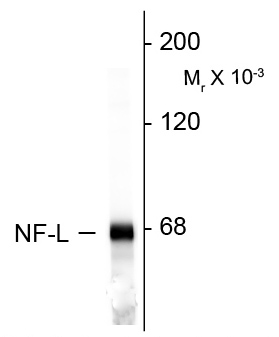
ARG52349 anti-Neurofilament NF-L antibody WB image
Western blot: Rat cortex lysate stained with ARG52349 anti-Neurofilament NF-L antibody.