ARG10732
anti-Neurofilament NF-L antibody
anti-Neurofilament NF-L antibody for ICC/IF,IHC-Frozen sections,Western blot and Human,Mouse,Rat
Controls and Markers antibody; Developmental Biology antibody; Neuroscience antibody; Signaling Transduction antibody; Neuronal Cytoskeletal antibody; Neurofilament antibody; Intermediate Neurofilament antibody
Overview
| Product Description | Rabbit Polyclonal antibody recognizes Neurofilament NF-L |
|---|---|
| Tested Reactivity | Hu, Ms, Rat |
| Predict Reactivity | Bov, Pig |
| Tested Application | ICC/IF, IHC-Fr, WB |
| Host | Rabbit |
| Clonality | Polyclonal |
| Isotype | IgG |
| Target Name | Neurofilament NF-L |
| Antigen Species | Human |
| Immunogen | Recombinant Human NF-L protein |
| Conjugation | Un-conjugated |
| Alternate Names | Neurofilament triplet L protein; 68 kDa neurofilament protein; CMT1F; NF68; NFL; CMT2E; Neurofilament light polypeptide; NF-L; PPP1R110 |
Application Instructions
| Application Suggestion |
|
||||||||
|---|---|---|---|---|---|---|---|---|---|
| Application Note | * The dilutions indicate recommended starting dilutions and the optimal dilutions or concentrations should be determined by the scientist. |
Properties
| Form | Liquid |
|---|---|
| Purification | Unpurified. |
| Buffer | Serum. |
| Storage Instruction | For continuous use, store undiluted antibody at 2-8°C for up to a week. For long-term storage, aliquot and store at -20°C or below. Storage in frost free freezers is not recommended. Avoid repeated freeze/thaw cycles. Suggest spin the vial prior to opening. The antibody solution should be gently mixed before use. |
| Note | For laboratory research only, not for drug, diagnostic or other use. |
Bioinformation
| Database Links | |
|---|---|
| Gene Symbol | NEFL |
| Gene Full Name | neurofilament, light polypeptide |
| Background | Neurofilaments are type IV intermediate filament heteropolymers composed of light, medium, and heavy chains. Neurofilaments comprise the axoskeleton and they functionally maintain the neuronal caliber. They may also play a role in intracellular transport to axons and dendrites. This gene encodes the light chain neurofilament protein. Mutations in this gene cause Charcot-Marie-Tooth disease types 1F (CMT1F) and 2E (CMT2E), disorders of the peripheral nervous system that are characterized by distinct neuropathies. A pseudogene has been identified on chromosome Y. [provided by RefSeq, Oct 2008] |
| Function | Neurofilaments usually contain three intermediate filament proteins: L, M, and H which are involved in the maintenance of neuronal caliber. [UniProt] |
| Research Area | Controls and Markers antibody; Developmental Biology antibody; Neuroscience antibody; Signaling Transduction antibody; Neuronal Cytoskeletal antibody; Neurofilament antibody; Intermediate Neurofilament antibody |
| Calculated MW | 62 kDa |
| PTM | O-glycosylated. Phosphorylated in the head and rod regions by the PKC kinase PKN1, leading to the inhibition of polymerization. Ubiquitinated in the presence of TRIM2 and UBE2D1. |
Images (4) Click the Picture to Zoom In
-
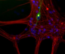
ARG10732 anti-Neurofilament NF-L antibody ICC/IF image
Immunocytochemistry: Mixed neuron / glia cultures from newborn Rat brain stained with monoclonal 7C5 to peripherin (green) and co-stained with ARG10732 anti-Neurofilament NF-L antibody (red). A class of large neurons, like the one in the middle of this image, contains peripherin, while the majority of neurons and their processes contain NF-L and not peripherin. Interestingly, the peripherin positive cells often contain a cytoplasmic inclusion next to the nucleus which stains for both peripherin and NF-L, and so appears golden in this image. The blue channel reveals the localization of DNA.
-
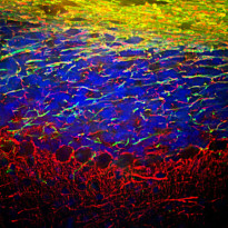
ARG10732 anti-Neurofilament NF-L antibody IHC-Fr image
Immunohistochemistry: Frozen section of Mouse cerebellum stained with ARG10732 anti-Neurofilament NF-L antibody (red) at 1:5000 dilution and costained with ARG52344 anti-Myelin Basic Protein antibody (green) at 1:5000 dilution. (Sample preparation: Following transcardial perfusion of Mouse with 4% paraformaldehyde, brain was post fixed for 24 hours, cut to 45 µM, and free-floating sections were stained with above antibodies.)
The NF-L antibody labels dendrites and axons of neuronal cells, and Myelin Basic Protein antibody stains myelin sheathes around axons.
-
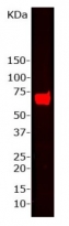
ARG10732 anti-Neurofilament NF-L antibody WB image
Western blot: Whole Rat brain homogenate stained with ARG10732 anti-Neurofilament NF-L antibody at 1:15000 dilution. A prominent band running with an apparent SDS-PAGE molecular weight of ~68 kDa corresponds to rodent NF-L.
-
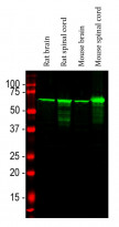
ARG10732 anti-Neurofilament NF-L antibody WB image
Western blot: Rat brain, Rat spinal cord, Mouse brain and Mouse spinal cord lysates stained with ARG10732 anti-Neurofilament NF-L antibody (green) at 1:20000 dilution.


