ARG52346
anti-Neurofilament NF-H phospho (KSP site) antibody [NAP4]
anti-Neurofilament NF-H phospho (KSP site) antibody [NAP4] for IHC-Formalin-fixed paraffin-embedded sections,Western blot and Mouse,Rat,Cow,Pig
Neuroscience antibody; Signaling Transduction antibody; Neurofilament antibody; Intermediate Neurofilament antibody
Overview
| Product Description | Mouse Monoclonal antibody [NAP4] recognizes Neurofilament NF-H when KSP site was phosphorylated |
|---|---|
| Tested Reactivity | Ms, Rat, Cow, Pig |
| Predict Reactivity | Hu |
| Tested Application | IHC-P, WB |
| Specificity | NAP4 recognizes phosphorylated NF-H KSP sequences but not non-phosphorylated KSP sequences. |
| Host | Mouse |
| Clonality | Monoclonal |
| Clone | NAP4 |
| Isotype | IgG1 |
| Target Name | Neurofilament NF-H |
| Antigen Species | Bovine |
| Immunogen | Purified bovine NF-H |
| Conjugation | Un-conjugated |
| Alternate Names | Neurofilament heavy polypeptide; 200 kDa neurofilament protein; NF-H; Neurofilament triplet H protein; NFH |
Application Instructions
| Application Suggestion |
|
||||||
|---|---|---|---|---|---|---|---|
| Application Note | Specific for the ~200k Neurofilament H protein. It recognizes phosphorylated NF-H KSP (lysine-serine-proline) type sequences. In some species there is some cross-reactivity with the related phosphorylated KSP sequences found in the related neurofilament subunit NF-M. It recognizes neurofilaments in frozen sections in tissue culture and in formalin fixed sections. * The dilutions indicate recommended starting dilutions and the optimal dilutions or concentrations should be determined by the scientist. |
Properties
| Form | Liquid |
|---|---|
| Purification | Total IgG fraction |
| Buffer | Total IgG fraction and 10 mM Sodium azide |
| Preservative | 10 mM Sodium azide |
| Storage Instruction | For continuous use, store undiluted antibody at 2-8°C for up to a week. For long-term storage, aliquot and store at -20°C or below. Storage in frost free freezers is not recommended. Avoid repeated freeze/thaw cycles. Suggest spin the vial prior to opening. The antibody solution should be gently mixed before use. |
| Note | For laboratory research only, not for drug, diagnostic or other use. |
Bioinformation
| Database Links | |
|---|---|
| Background | Neurofilaments are the 10nm or intermediate filament proteins found specifically in neurons, and are composed predominantly of three major proteins called NF-L, NF-M and NF-H . NF-H is the neurofilament high or heavy molecular weight polypeptide and runs on SDS-PAGE gels at 200-220 kDa, with some variability across species boundaries. Antibodies to NF-H are useful for identifying neuronal cells and their processes in tissue sections and in tissue culture. NF-H antibodies can also be useful to visualize neurofilament accumulations seen in many neurological diseases, such as Amyotrophic Lateral Sclerosis (Lou Gehrig's disease) (2) and Alzheimer's disease . |
| Research Area | Neuroscience antibody; Signaling Transduction antibody; Neurofilament antibody; Intermediate Neurofilament antibody |
| Calculated MW | 112 kDa |
| PTM | There are a number of repeats of the tripeptide K-S-P, NFH is phosphorylated on a number of the serines in this motif. It is thought that phosphorylation of NFH results in the formation of interfilament cross bridges that are important in the maintenance of axonal caliber. Phosphorylation seems to play a major role in the functioning of the larger neurofilament polypeptides (NF-M and NF-H), the levels of phosphorylation being altered developmentally and coincidentally with a change in the neurofilament function. Phosphorylated in the head and rod regions by the PKC kinase PKN1, leading to the inhibition of polymerization. |
Images (3) Click the Picture to Zoom In
-

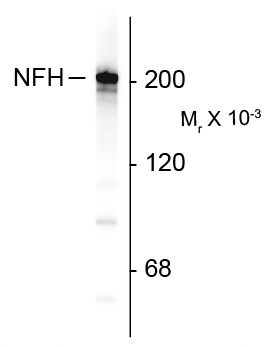
ARG52346 anti-Neurofilament NF-H antibody [NAP4] WB image
Western Blot: Rat cortex lysate showing specific immunolabeling of the ~200k NF-H protein stained with ARG52346 anti-Neurofilament NF-H antibody [NAP4].
-
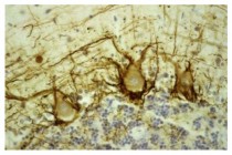
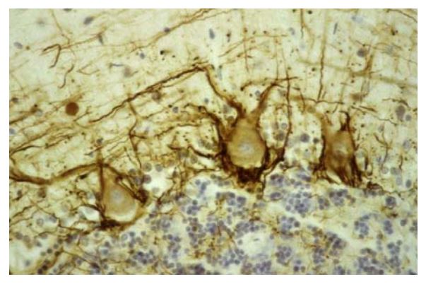
ARG52346 anti-Neurofilament NF-H antibody [NAP4] IHC image
Immunohistochemistry: Human cerebellar cortex stained with ARG52346 anti-Neurofilament NF-H antibody [NAP4] showing labeling of NF-H (brown) in basket cell axons surrounding the large Purkinje neurons.
-
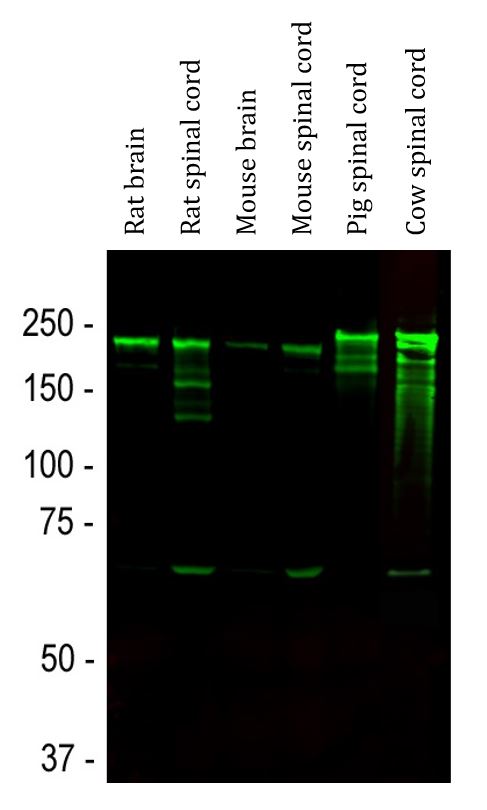
ARG52346 anti-Neurofilament NF-H antibody [NAP4] WB image
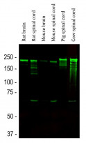
Western blot: Rat brain, Rat spinal cord, Mouse brain, Mouse spinal cord, Pig spinal cord and Cow spinal cord lysates stained with ARG52346 anti-Neurofilament NF-H antibody [NAP4] (green) at 1:10000 dilution.
Strong band at about 200-220 kDa corresponds to the major phosphorylated form of the NF-H subunit. A minor band at about 160 kDa is the non-phosphorylated NF-H. Smaller proteolytic fragments of NF-H are also detected in spinal cord preparations.
Clone References
Compartmentation of alpha-internexin and neurofilament triplet proteins in cultured hippocampal neurons.
WB / Rat


