ARG10761
anti-Neurofilament NF-H antibody
anti-Neurofilament NF-H antibody for ICC/IF,IHC-Frozen sections,Western blot and Mouse,Rat
Neuroscience antibody; Signaling Transduction antibody; Neurofilament antibody; Intermediate Neurofilament antibody
Overview
| Product Description | Rabbit Polyclonal antibody recognizes Neurofilament NF-H |
|---|---|
| Tested Reactivity | Ms, Rat |
| Predict Reactivity | Hu, Bov, Hrs, Pig |
| Tested Application | ICC/IF, IHC-Fr, WB |
| Specificity | This antibody reacts very strongly with NF-H KSP phosphorylated repeats. Reactivity with non-phosphorylated KSP sequences is orders of magnitude weaker. |
| Host | Rabbit |
| Clonality | Polyclonal |
| Isotype | IgG |
| Target Name | Neurofilament NF-H |
| Antigen Species | Bovine |
| Immunogen | Native NF-H purified from Bovine spinal cord. |
| Conjugation | Un-conjugated |
| Alternate Names | Neurofilament heavy polypeptide; 200 kDa neurofilament protein; NF-H; Neurofilament triplet H protein; NFH |
Application Instructions
| Application Suggestion |
|
||||||||
|---|---|---|---|---|---|---|---|---|---|
| Application Note | * The dilutions indicate recommended starting dilutions and the optimal dilutions or concentrations should be determined by the scientist. |
Properties
| Form | Liquid |
|---|---|
| Purification | Unpurified. |
| Buffer | Serum. |
| Storage Instruction | For continuous use, store undiluted antibody at 2-8°C for up to a week. For long-term storage, aliquot and store at -20°C or below. Storage in frost free freezers is not recommended. Avoid repeated freeze/thaw cycles. Suggest spin the vial prior to opening. The antibody solution should be gently mixed before use. |
| Note | For laboratory research only, not for drug, diagnostic or other use. |
Bioinformation
| Database Links | |
|---|---|
| Gene Symbol | NEFH |
| Gene Full Name | neurofilament, heavy polypeptide |
| Background | Neurofilaments are type IV intermediate filament heteropolymers composed of light, medium, and heavy chains. Neurofilaments comprise the axoskeleton and functionally maintain neuronal caliber. They may also play a role in intracellular transport to axons and dendrites. This gene encodes the heavy neurofilament protein. This protein is commonly used as a biomarker of neuronal damage and susceptibility to amyotrophic lateral sclerosis (ALS) has been associated with mutations in this gene. [provided by RefSeq, Oct 2008] |
| Function | Neurofilaments usually contain three intermediate filament proteins: L, M, and H which are involved in the maintenance of neuronal caliber. NF-H has an important function in mature axons that is not subserved by the two smaller NF proteins. [UniProt] |
| Research Area | Neuroscience antibody; Signaling Transduction antibody; Neurofilament antibody; Intermediate Neurofilament antibody |
| Calculated MW | 112 kDa |
| PTM | There are a number of repeats of the tripeptide K-S-P, NFH is phosphorylated on a number of the serines in this motif. It is thought that phosphorylation of NFH results in the formation of interfilament cross bridges that are important in the maintenance of axonal caliber. Phosphorylation seems to play a major role in the functioning of the larger neurofilament polypeptides (NF-M and NF-H), the levels of phosphorylation being altered developmentally and coincidentally with a change in the neurofilament function. Phosphorylated in the head and rod regions by the PKC kinase PKN1, leading to the inhibition of polymerization. |
Images (4) Click the Picture to Zoom In
-
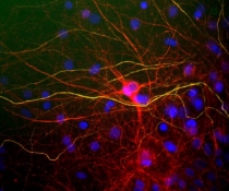
ARG10761 anti-Neurofilament NF-H antibody ICC/IF image
Immunocytochemistry: Rat Mixed neuron / glia cultures stained with ARG10761 anti-Neurofilament NF-H antibody (green) and co-stained with chicken antibody to neurofilament subunit NF-L (red). Axons contain phosphorylated NF-H and NF-L so appear yellow, while dendrites and perikarya only contain NF-L and so appear red. DNA is shown in blue.
-
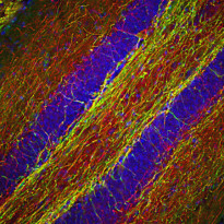
ARG10761 anti-Neurofilament NF-H antibody IHC-Fr image
Immunohistochemistry: Frozen section of Mouse hippocampus stained with ARG10761 anti-Neurofilament NF-H antibody (red) at 1:2000 dilution and costained with ARG10723 anti-Myelin Basic Protein antibody [7G7] (green) at 1:5000 dilution. DAPI (blue) for nuclear staining. (Sample preparation: Following transcardial perfusion with 4% paraformaldehyde, brain was post fixed for 24 hours, cut to 45 µM, and free-floating sections were stained with above antibodies.)
The NF-H antibody labels a network of axons of different neurons, while the Myelin Basic Protein antibody stains myelin sheath around these axons.
-

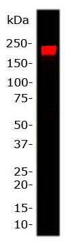
ARG10761 anti-Neurofilament NF-H antibody WB image
Western blot: 20 µg of Rat brain lysate stained with ARG10761 anti-Neurofilament NF-H antibody at 1:25,000 dilution. A prominent band at 200 kDa corresponds to phosphorylated form of NF-H.
-
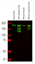
ARG10761 anti-Neurofilament NF-H antibody WB image
Western blot: Rat brain, Rat spinal cord, Mouse brain and Mouse spinal cord lysates stained with ARG10761 anti-Neurofilament NF-H antibody (green) at 1:10000 dilution.
Strong band at about 220 kDa corresponds to the phosphorylated axonal form of the NF-H subunit. Smaller proteolytic fragments of NF-H are also detected.