ARG44113
anti-NUMA1 antibody
anti-NUMA1 antibody for Flow cytometry,ICC/IF,IHC-Formalin-fixed paraffin-embedded sections,Western blot and Human,Mouse,Rat,Monkey
Overview
| Product Description | Rabbit Polyclonal recognizes NUMA1 |
|---|---|
| Tested Reactivity | Hu, Ms, Rat, Mk |
| Tested Application | FACS, ICC/IF, IHC-P, WB |
| Host | Rabbit |
| Clonality | Polyclonal |
| Isotype | IgG |
| Target Name | NUMA1 |
| Antigen Species | Human |
| Immunogen | Human NUMA1 recombinant protein (Position: M1-E1954). |
| Conjugation | Un-conjugated |
| Alternate Names | NUMA1; Nuclear Mitotic Apparatus Protein 1; NUMA; Nuclear Matrix Protein-22; SP-H Antigen; NMP-22; Centrophilin Stabilizes Mitotic Spindle In Mitotic Cells; Nuclear Mitotic Apparatus Protein; Structural Nuclear Protein; NuMA Protein; NMP22 |
Application Instructions
| Application Suggestion |
|
||||||||||
|---|---|---|---|---|---|---|---|---|---|---|---|
| Application Note | The dilutions indicate recommended starting dilutions and the optimal dilutions or concentrations should be determined by the scientist. |
Properties
| Form | Liquid |
|---|---|
| Purification | Affinity purification with immunogen. |
| Buffer | 0.9% NaCl, 0.2% Na2HPO4, 0.05% Sodium azide and 4% Trehalose. |
| Preservative | 0.05% Sodium azide |
| Stabilizer | 4% Trehalose |
| Concentration | 0.5 mg/ml |
| Storage Instruction | For continuous use, store undiluted antibody at 2-8°C for up to a week. For long-term storage, aliquot and store at -20°C or below. Storage in frost free freezers is not recommended. Avoid repeated freeze/thaw cycles. Suggest spin the vial prior to opening. The antibody solution should be gently mixed before use. |
| Note | For laboratory research only, not for drug, diagnostic or other use. |
Bioinformation
| Database Links |
Swiss-port # Q14980 Human Nuclear mitotic apparatus protein 1 |
|---|---|
| Gene Symbol | NUMA1 |
| Gene Full Name | Nuclear Mitotic Apparatus Protein 1 |
| Background | This gene encodes a large protein that forms a structural component of the nuclear matrix. The encoded protein interacts with microtubules and plays a role in the formation and organization of the mitotic spindle during cell division. Chromosomal translocation of this gene with the RARA (retinoic acid receptor, alpha) gene on chromosome 17 have been detected in patients with acute promyelocytic leukemia. Alternative splicing results in multiple transcript variants. |
| Function | Microtubule (MT)-binding protein that plays a role in the formation and maintenance of the spindle poles and the alignement and the segregation of chromosomes during mitotic cell division. |
| Cellular Localization | Cell membrane, Chromosome, Cytoplasm, Cytoskeleton, Membrane, Microtubule, Nucleus |
| Calculated MW | 238 kDa |
| PTM | Acetylation, ADP-ribosylation, Glycoprotein, Isopeptide bond, Lipoprotein, Phosphoprotein, Ubl conjugation |
Images (8) Click the Picture to Zoom In
-
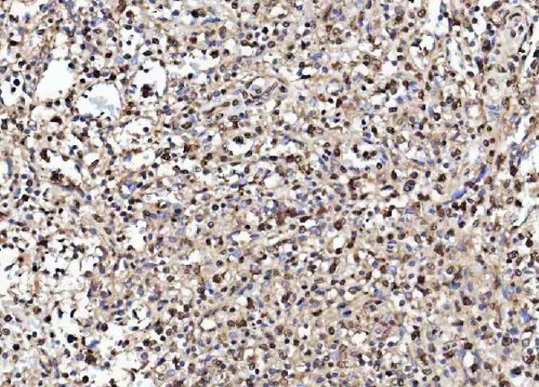
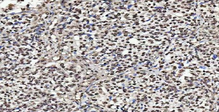
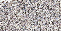
ARG44113 anti-NUMA1 antibody IHC-P image
Immunohistochemistry: Human acinar adenocarcinoma of prostate stained with ARG44113 anti-NUMA1 antibody at 2 μg/ml dilution.
-

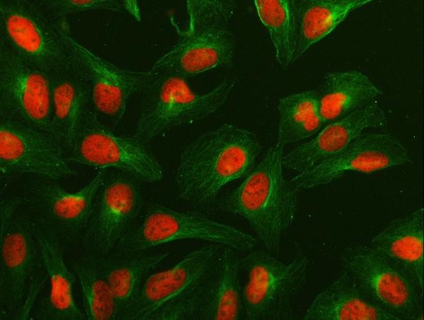
ARG44113 anti-NUMA1 antibody ICC/IF image
Immunofluorescence: U2OS stained with ARG44113 anti-NUMA1 antibody at 5 μg/ml dilution.
-
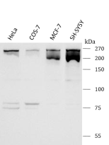
ARG44113 anti-NUMA1 antibody WB image
Western blot: Hela, COS-7, MCF-7 and SH-SY5Y stained with ARG44113 anti-NUMA1 antibody at 0.5 μg/ml dilution.
-
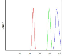
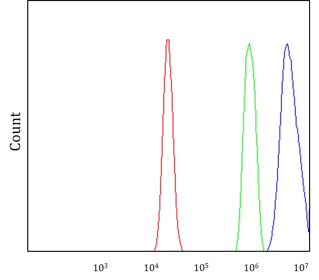
ARG44113 anti-NUMA1 antibody FACS image
Flow Cytometry: RT4 stained with ARG44113 anti-NUMA1 antibody at 1 µg/10^6 cells dilution.
-
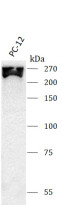
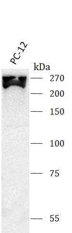
ARG44113 anti-NUMA1 antibody WB image
Western blot: PC-12 stained with ARG44113 anti-NUMA1 antibody at 0.5 μg/ml dilution.
-
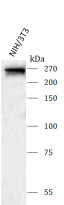
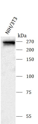
ARG44113 anti-NUMA1 antibody WB image
Western blot: NIH/3T3 stained with ARG44113 anti-NUMA1 antibody at 0.5 μg/ml dilution.
-
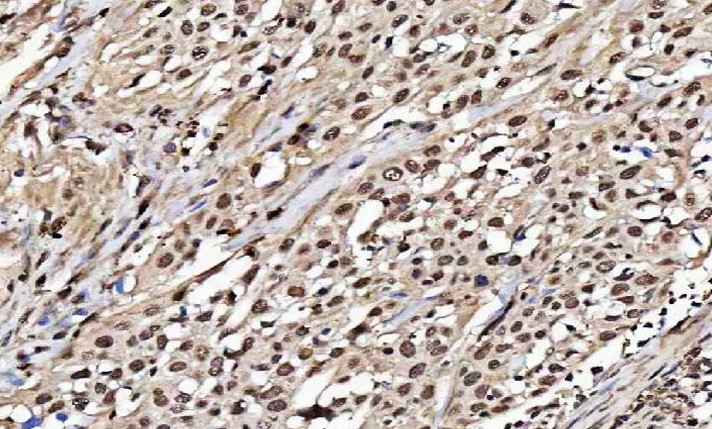
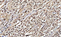
ARG44113 anti-NUMA1 antibody IHC-P image
Immunohistochemistry: Human breast cancer stained with ARG44113 anti-NUMA1 antibody at 2 μg/ml dilution.
-
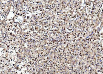
ARG44113 anti-NUMA1 antibody IHC-P image
Immunohistochemistry: Human esophageal squamous carcinoma stained with ARG44113 anti-NUMA1 antibody at 2 μg/ml dilution.