ARG52377
anti-NSE / Neuron Specific Enolase antibody
anti-NSE / Neuron Specific Enolase antibody for ICC/IF,Western blot and Human,Mouse,Rat
Cancer antibody; Gene Regulation antibody; Metabolism antibody; Signaling Transduction antibody

Overview
| Product Description | Rabbit Polyclonal antibody recognizes NSE / Neuron Specific Enolase |
|---|---|
| Tested Reactivity | Hu, Ms, Rat |
| Tested Application | ICC/IF, WB |
| Host | Rabbit |
| Clonality | Polyclonal |
| Isotype | IgG |
| Target Name | NSE / Neuron Specific Enolase |
| Antigen Species | Human |
| Immunogen | Recombinant human NSE expressed in and purified from E. coli |
| Conjugation | Un-conjugated |
| Alternate Names | Neural enolase; NSE; Enolase 2; Gamma-enolase; 2-phospho-D-glycerate hydro-lyase; HEL-S-279; Neuron-specific enolase; EC 4.2.1.11 |
Application Instructions
| Application Suggestion |
|
||||||
|---|---|---|---|---|---|---|---|
| Application Note | Specific for the ~47kDa NSE protein. * The dilutions indicate recommended starting dilutions and the optimal dilutions or concentrations should be determined by the scientist. |
Properties
| Form | Liquid |
|---|---|
| Purification | Neat Serum |
| Buffer | Neat serum |
| Storage Instruction | For continuous use, store undiluted antibody at 2-8°C for up to a week. For long-term storage, aliquot and store at -20°C or below. Storage in frost free freezers is not recommended. Avoid repeated freeze/thaw cycles. Suggest spin the vial prior to opening. The antibody solution should be gently mixed before use. |
| Note | For laboratory research only, not for drug, diagnostic or other use. |
Bioinformation
| Database Links | |
|---|---|
| Gene Symbol | ENO2 |
| Gene Full Name | enolase 2 (gamma, neuronal) |
| Background | Neuron specific enolase (NSE) is an enzyme which catalyzes the conversion of 2-phosphoglycerate to phosphoenolpyruvate in the glycolytic pathway, and also the reverse reaction in gluconeogenesis. It is one of three mammalian enolases, which are also known as ENO1, ENO2, and ENO3 or alternately as enolase alpha, beta and gamma. The three enolases have different cell type specific expression patterns, so that antibodies to them are useful cell type specific markers.(MacAlesse et al., 1988). NSE corresponds to ENO2 or enolase gamma and is heavily expressed in neuronal cells. ENO1 is also known as enolase alpha and as non-neuronal enolase. The third enolase, ENO3 or enolase beta, is expressed in muscle cells. Since neurons require a great deal of energy, they are very rich in glycolytic enzymes such a GAPDH and NSE. Antibodies to this protein are therefore useful to identify neuronal cell bodies, developing neuronal lineage and neuroendocrine cells. Release of NSE from damaged neurons into CSF and blood has also been used as a biomarker of neuronal injury . |
| Highlight | Related products: NSE antibodies; Anti-Rabbit IgG secondary antibodies; Related news: Neuronal Development Marker |
| Research Area | Cancer antibody; Gene Regulation antibody; Metabolism antibody; Signaling Transduction antibody |
| Calculated MW | 47 kDa |
Images (5) Click the Picture to Zoom In
-
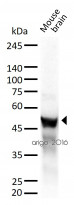
ARG52377 anti-NSE / Neuron Specific Enolase antibody WB image
Western blot: 30 µg of Mouse brain lysate stained with ARG52377 anti-NSE / Neuron Specific Enolase antibody at 1:2000 dilution.
-
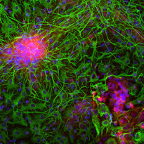
ARG52377 anti-NSE / Neuron Specific Enolase antibody ICC/IF image
Immunofluorescence: Mixed cortical neuron-glial cell culture from E20 Rat stained with ARG52377 anti-NSE / Neuron Specific Enolase antibody (red) at 1:500 dilution, and costained with anti-GFAP antibody (green) at 1:5000 dilution. Hoechst (blue) for nuclear staining.
The NSE antibody labels protein expressed in neuronal cells, while the GFAP antibody stains intermediate filaments in astrocytic and certain other glial cells.
-
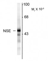
ARG52377 anti-NSE / Neuron Specific Enolase antibody WB image
Western blot: Rat cortex homogenate showing specific immunolabeling of the ~ 47k NSE protein stained with ARG52377 anti-NSE / Neuron Specific Enolase antibody.
-
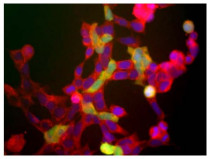
ARG52377 anti-NSE / Neuron Specific Enolase antibody ICC/IF image
Immunofluorescence: HEK 293 cells showing staining with ARG52377 anti-NSE / Neuron Specific Enolase antibody (red). The green channels shows staining with ARG52465 anti-UCHL1 antibody [BH7].
-
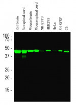
ARG52377 anti-NSE / Neuron Specific Enolase antibody WB image
Western blot: Rat brain, Rat spinal cord, Mouse brain, Mouse spinal cord, NIH/3T3, HEK293, HeLa, SH-SY5Y and C6 cell lysates stained with ARG52377 anti-NSE / Neuron Specific Enolase antibody (green) at 1:5000 dilution.
A single band at about 47 kDa corresponds to the NSE protein, seen only in extracts containing neurons or neuronal lineage cells.


Customer's Feedback
Excellent

Review for anti-NSE / Neuron Specific Enolase antibody
Application:WB
Sample:Rat brain
Sample Loading Amount:30 µg
Primary Antibody Dilution Factor:1:2000
Primary Antibody Incubation Time:overnight
Primary Antibody Incubation Temperature:4 ºC

Excellent

Review for anti-NSE / Neuron Specific Enolase antibody
Application:WB
Sample:U87-MG
Sample Loading Amount:30 µg
Primary Antibody Dilution Factor:1:2000
Primary Antibody Incubation Time:overnight
Primary Antibody Incubation Temperature:4 ºC


