ARG40530
anti-NPHP1 / Nephrocystin 1 antibody
anti-NPHP1 / Nephrocystin 1 antibody for ICC/IF,IHC-Formalin-fixed paraffin-embedded sections,Western blot and Human,Mouse,Rat
Overview
| Product Description | Rabbit Polyclonal antibody recognizes NPHP1 / Nephrocystin 1 |
|---|---|
| Tested Reactivity | Hu, Ms, Rat |
| Tested Application | ICC/IF, IHC-P, WB |
| Host | Rabbit |
| Clonality | Polyclonal |
| Isotype | IgG |
| Target Name | NPHP1 / Nephrocystin 1 |
| Antigen Species | Human |
| Immunogen | Recombinant fusion protein corresponding to aa. 345-614 of Human NPHP1 / Nephrocystin 1 (NP_001121651.1). |
| Conjugation | Un-conjugated |
| Alternate Names | JBTS4; SLSN1; Juvenile nephronophthisis 1 protein; Nephrocystin-1; NPH1 |
Application Instructions
| Application Suggestion |
|
||||||||
|---|---|---|---|---|---|---|---|---|---|
| Application Note | * The dilutions indicate recommended starting dilutions and the optimal dilutions or concentrations should be determined by the scientist. | ||||||||
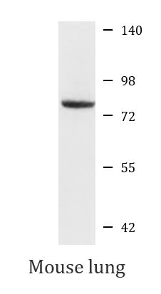
| Positive Control | Mouse lung | ||||||||
| Observed Size | 80 kDa |
Properties
| Form | Liquid |
|---|---|
| Purification | Affinity purified. |
| Buffer | PBS (pH 7.3), 0.02% Sodium azide and 50% Glycerol. |
| Preservative | 0.02% Sodium azide |
| Stabilizer | 50% Glycerol |
| Storage Instruction | For continuous use, store undiluted antibody at 2-8°C for up to a week. For long-term storage, aliquot and store at -20°C. Storage in frost free freezers is not recommended. Avoid repeated freeze/thaw cycles. Suggest spin the vial prior to opening. The antibody solution should be gently mixed before use. |
| Note | For laboratory research only, not for drug, diagnostic or other use. |
Bioinformation
| Database Links | |
|---|---|
| Gene Symbol | NPHP1 |
| Gene Full Name | nephronophthisis 1 (juvenile) |
| Background | This gene encodes a protein with src homology domain 3 (SH3) patterns. This protein interacts with Crk-associated substrate, and it appears to function in the control of cell division, as well as in cell-cell and cell-matrix adhesion signaling, likely as part of a multifunctional complex localized in actin- and microtubule-based structures. Mutations in this gene cause familial juvenile nephronophthisis type 1, a kidney disorder involving both tubules and glomeruli. Defects in this gene are also associated with Senior-Loken syndrome type 1, also referred to as juvenile nephronophthisis with Leber amaurosis, which is characterized by kidney and eye disease, and with Joubert syndrome type 4, which is characterized by cerebellar ataxia, oculomotor apraxia, psychomotor delay and neonatal breathing abnormalities, sometimes including retinal dystrophy and renal disease. Multiple transcript variants encoding different isoforms have been found for this gene. [provided by RefSeq, Jul 2008] |
| Function | Together with BCAR1 it may play a role in the control of epithelial cell polarity. Involved in the organization of apical junctions in kidney cells together with NPHP4 and RPGRIP1L/NPHP8 (By similarity). Does not seem to be strictly required for ciliogenesis (By similarity). Seems to help to recruit PTK2B/PYK2 to cell matrix adhesions, thereby initiating phosphorylation of PTK2B/PYK2 and PTK2B/PYK2-dependent signaling. May play a role in the regulation of intraflagellar transport (IFT) during cilia assembly. Required for normal retina development. In connecting photoreceptor cilia influences the movement of some IFT proteins such as IFT88 and WDR19. Involved in spermatogenesis (By similarity). [UniProt] |
| Cellular Localization | Cell junction, adherens junction, tight junction. Cell projection, cilium. Cytoplasm, cytoskeleton, cilium axoneme. Note=Colocalizes with E-cadherin and BCAR1 at or near the cell-cell adherens junctions. Localized to respiratory cilia axoneme. Localized to the transition zone of respiratory cilia, photoreceptor-connecting cilia and renal monocilia. [UniProt] |
| Calculated MW | 83 kDa |
| PTM | Phosphorylation by CK2 is required for the interaction with PACS1 and the targeting to the base region of cilia. [UniProt] |
Images (2) Click the Picture to Zoom In
-

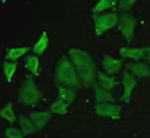
ARG40530 anti-NPHP1 / Nephrocystin 1 antibody ICC/IF image
Immunofluorescence: MCF7 cells stained with ARG40530 anti-NPHP1 / Nephrocystin 1 antibody.
-

ARG40530 anti-NPHP1 / Nephrocystin 1 antibody WB image
Western blot: 25 µg of Mouse lung lysate stained with ARG40530 anti-NPHP1 / Nephrocystin 1 antibody at 1:1000 dilution.