ARG64942
anti-NOXA antibody
anti-NOXA antibody for Western blot and Human
Cancer antibody; Cell Biology and Cellular Response antibody; Cell Death antibody; Metabolism antibody
Overview
| Product Description | Goat Polyclonal antibody recognizes NOXA |
|---|---|
| Tested Reactivity | Hu |
| Tested Application | WB |
| Host | Goat |
| Clonality | Polyclonal |
| Isotype | IgG |
| Target Name | NOXA |
| Antigen Species | Human |
| Immunogen | C-TQLRRFGDKLNFRQK |
| Conjugation | Un-conjugated |
| Alternate Names | NOXA; APR; Phorbol-12-myristate-13-acetate-induced protein 1; PMA-induced protein 1; Immediate-early-response protein APR; Protein Noxa |
Application Instructions
| Application Suggestion |
|
||||
|---|---|---|---|---|---|
| Application Note | WB: Recommend incubate at RT for 1h. * The dilutions indicate recommended starting dilutions and the optimal dilutions or concentrations should be determined by the scientist. |
Properties
| Form | Liquid |
|---|---|
| Purification | Purified from goat serum by antigen affinity chromatography. |
| Buffer | Tris saline (pH 7.3), 0.02% Sodium azide and 0.5% BSA. |
| Preservative | 0.02% Sodium azide |
| Stabilizer | 0.5% BSA |
| Concentration | 0.5 mg/ml |
| Storage Instruction | For continuous use, store undiluted antibody at 2-8°C for up to a week. For long-term storage, aliquot and store at -20°C or below. Storage in frost free freezers is not recommended. Avoid repeated freeze/thaw cycles. Suggest spin the vial prior to opening. The antibody solution should be gently mixed before use. |
| Note | For laboratory research only, not for drug, diagnostic or other use. |
Bioinformation
| Database Links |
Swiss-port # Q13794 Human Phorbol-12-myristate-13-acetate-induced protein 1 |
|---|---|
| Gene Symbol | PMAIP1 |
| Gene Full Name | phorbol-12-myristate-13-acetate-induced protein 1 |
| Function | Promotes activation of caspases and apoptosis. Promotes mitochondrial membrane changes and efflux of apoptogenic proteins from the mitochondria. Contributes to p53/TP53-dependent apoptosis after radiation exposure. Promotes proteasomal degradation of MCL1. Competes with BAK1 for binding to MCL1 and can displace BAK1 from its binding site on MCL1 (By similarity). Competes with BIM/BCL2L11 for binding to MCL1 and can displace BIM/BCL2L11 from its binding site on MCL1. [UniProt] |
| Research Area | Cancer antibody; Cell Biology and Cellular Response antibody; Cell Death antibody; Metabolism antibody |
| Calculated MW | 6 kDa |
Images (1) Click the Picture to Zoom In
-
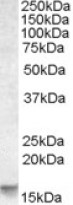
ARG64942 anti-NOXA antibody WB image
Western Blot: Human Cerebellum lysate (35 µg protein in RIPA buffer) stained with ARG64942 anti-NOXA antibody at 0.03 µg/ml dilution.


