ARG52368
anti-NMDAR2B phospho (Tyr1336) antibody
anti-NMDAR2B phospho (Tyr1336) antibody for IHC-Formalin-fixed paraffin-embedded sections,Western blot and Rat
Neuroscience antibody; Postsynaptic Receptor antibody
Overview
| Product Description | Rabbit Polyclonal antibody recognizes NMDAR2B phospho (Tyr1336) |
|---|---|
| Tested Reactivity | Rat |
| Predict Reactivity | Hu, Ms, NHuPrm |
| Tested Application | IHC-P, WB |
| Host | Rabbit |
| Clonality | Polyclonal |
| Isotype | IgG |
| Target Name | NMDAR2B |
| Antigen Species | Rat |
| Immunogen | Synthetic phospho-peptide corresponding to amino acid residues surrounding Tyr1336 conjugated to KLH |
| Conjugation | Un-conjugated |
| Alternate Names | MRD6; EIEE27; NR2B; hNR3; GluN2B; NR3; N-methyl D-aspartate receptor subtype 2B; Glutamate receptor ionotropic, NMDA 2B; Glutamate [NMDA] receptor subunit epsilon-2; N-methyl-D-aspartate receptor subunit 3; NMDAR2B |
Application Instructions
| Application Suggestion |
|
||||||
|---|---|---|---|---|---|---|---|
| Application Note | Specific for ~180k NMDAR NR2B subunit protein phosphorylated at Tyr1336. Immunolabeling of the NMDAR NR2B subunit band is blocked by λ-phosphatase treatment. * The dilutions indicate recommended starting dilutions and the optimal dilutions or concentrations should be determined by the scientist. |
Properties
| Form | Liquid |
|---|---|
| Purification | Affinity Purified |
| Buffer | 10 mM HEPES (pH 7.5), 150 mM NaCl, 0.1 mg/ml BSA and 50% Glycerol |
| Stabilizer | 0.1 mg/ml BSA, 50% Glycerol |
| Storage Instruction | For continuous use, store undiluted antibody at 2-8°C for up to a week. For long-term storage, aliquot and store at -20°C. Storage in frost free freezers is not recommended. Avoid repeated freeze/thaw cycles. Suggest spin the vial prior to opening. The antibody solution should be gently mixed before use. |
| Note | For laboratory research only, not for drug, diagnostic or other use. |
Bioinformation
| Database Links |
Swiss-port # Q00960 Rat Glutamate receptor ionotropic, NMDA 2B |
|---|---|
| Gene Symbol | GRIN2B |
| Gene Full Name | glutamate receptor, ionotropic, N-methyl D-aspartate 2B |
| Background | The NMDAR plays an essential role in memory, neuronal development and it has also been implicated in several disorders of the central nervous system including Alzheimer’s, epilepsy and ischemic neuronal cell death (Grosshans et al., 2002; Wenthold et al., 2003; Carroll and Zukin, 2002). The rat NMDAR1 (NR1) was the first subunit of the NMDAR to be cloned. The NR1 protein can form NMDA activated channels when expressed in Xenopus oocytes but the currents in such channels are much smaller than those seen in situ. Channels with more physiological characteristics are produced when the NR1 subunit is combined with one or more of the NMDAR2 (NR2 A-D) subunits (Ishii et al., 1993). Phosphorylation of Tyr1336 is thought to potentiate NMDA receptor-dependent influx of calcium (Takasu et al., 2002) and ischemia may also increase the phosphorylation of this site (Takagi et al., 2003). |
| Research Area | Neuroscience antibody; Postsynaptic Receptor antibody |
| Calculated MW | 166 kDa |
| PTM | Phosphorylation at Ser-1303 by DAPK1 enhances synaptic NMDA receptor channel activity. |
Images (1) Click the Picture to Zoom In
-
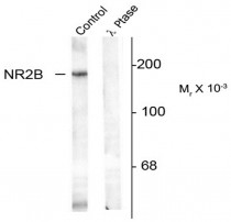
ARG52368 anti-NMDAR2B phospho (Tyr1336) antibody WB image
Western blot: Rat hippocampal lysate showing specific immunolabeling of the ~180k NR2B subunit of the NMDAR (Control) stained with ARG52368 anti-NMDAR2B phospho (Tyr1336) antibody. The phosphospecificty of this labeling is shown in the second lane (lambda-phosphatase: lambda Ptase). The blot is identical to the control except that it was incubated in lambda Ptase (1200 units for 30 min) before being exposed to the Anti-Phospho-Tyr1336 NMDA NR2B subunit. The immunolabeling of NR2B is completely eliminated by treatment with lambda Ptase.


