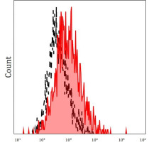
ARG43867
anti-NG2 / Chondroitin sulfate proteoglycan 4 antibody [7.1] (APC)
anti-NG2 / Chondroitin sulfate proteoglycan 4 antibody [7.1] (APC) for Flow cytometry and Human
Angiogenesis Study antibody; Mural cell Marker antibody
Overview
| Product Description | APC-conjugated Mouse Monoclonal antibody recognize NG2 / Chondroitin sulfate proteoglycan 4. |
|---|---|
| Tested Reactivity | Hu |
| Tested Application | FACS |
| Host | Mouse |
| Clonality | Monoclonal |
| Clone | 7.1 |
| Isotype | IgG1 |
| Target Name | NG2 / Chondroitin sulfate proteoglycan 4 |
| Antigen Species | Human |
| Immunogen | Human BMSCs infected with NG2 / Chondroitin sulfate proteoglycan 4 |
| Conjugation | APC |
| Protein Full Name | Chondroitin sulfate proteoglycan 4 |
| Alternate Names | HMW-MAA; MCSPG; Chondroitin sulfate proteoglycan NG2; MCSP; Chondroitin sulfate proteoglycan 4; MSK16; NG2; Melanoma chondroitin sulfate proteoglycan; MEL-CSPG; Melanoma-associated chondroitin sulfate proteoglycan |
Application Instructions
| Application Suggestion |
|
||||
|---|---|---|---|---|---|
| Application Note | * The dilutions indicate recommended starting dilutions and the optimal dilutions or concentrations should be determined by the scientist. |
Properties
| Form | Liquid |
|---|---|
| Purification | Purified |
| Buffer | PBS (pH 7.4) and 15 mM Sodium azide |
| Preservative | 15 mM Sodium azide |
| Storage Instruction | Aliquot and store in the dark at 4°C. Keep protected from prolonged exposure to light. Do not freeze. Suggest spin the vial prior to opening. The antibody solution should be gently mixed before use. |
Bioinformation
| Database Links |
Swiss-port # Q6UVK1 Human Chondroitin sulfate proteoglycan 4 |
|---|---|
| Gene Symbol | CSPG4 |
| Gene Full Name | Chondroitin Sulfate Proteoglycan 4 |
| Background | Chondroitin sulfate proteoglycan 4 (CSPG4): A human melanoma-associated chondroitin sulfate proteoglycan plays a role in stabilizing cell-substratum interactions during early events of melanoma cell spreading on endothelial basement membranes. CSPG4 represents an integral membrane chondroitin sulfate proteoglycan expressed by human malignant melanoma cells. [provided by RefSeq, Jul 2008] |
| Function | Proteoglycan playing a role in cell proliferation and migration which stimulates endothelial cells motility during microvascular morphogenesis. May also inhibit neurite outgrowth and growth cone collapse during axon regeneration. Cell surface receptor for collagen alpha 2(VI) which may confer cells ability to migrate on that substrate. Binds through its extracellular N-terminus growth factors, extracellular matrix proteases modulating their activity. May regulate MPP16-dependent degradation and invasion of type I collagen participating in melanoma cells invasion properties. May modulate the plasminogen system by enhancing plasminogen activation and inhibiting angiostatin. Functions also as a signal transducing protein by binding through its cytoplasmic C-terminus scaffolding and signaling proteins. May promote retraction fiber formation and cell polarization through Rho GTPase activation. May stimulate alpha-4, beta-1 integrin-mediated adhesion and spreading by recruiting and activating a signaling cascade through CDC42, ACK1 and BCAR1. May activate FAK and ERK1/ERK2 signaling cascades. [UniProt] |
| Cellular Localization | Cell membrane; Single-pass type I membrane protein; Extracellular side. Apical cell membrane; Single-pass type I membrane protein; Extracellular side. Cell projection, lamellipodium membrane; Single-pass type I membrane protein; Extracellular side. Cell surface. Note=Localized at the apical plasma membrane it relocalizes to the lamellipodia of astrocytoma upon phosphorylation by PRKCA. Localizes to the retraction fibers. Localizes to the plasma membrane of oligodendrocytes. [UniProt] |
| Research Area | Angiogenesis Study antibody; Mural cell Marker antibody |
| Calculated MW | 251 kDa |
| PTM | O-glycosylated; contains glycosaminoglycan chondroitin sulfate which are required for proper localization and function in stress fiber formation (By similarity). Involved in interaction with MMP16 and ITGA4. Phosphorylation by PRKCA regulates its subcellular location and function in cell motility. [UniProt] |
Images (1) Click the Picture to Zoom In


