ARG43375
anti-NFATc2 / NFAT1 phospho (Ser326) antibody
anti-NFATc2 / NFAT1 phospho (Ser326) antibody for Western blot and Human,Mouse,Rat
Overview
| Product Description | Rabbit Polyclonal antibody recognizes NFATc2 / NFAT1 phospho (Ser326) |
|---|---|
| Tested Reactivity | Hu, Ms, Rat |
| Tested Application | WB |
| Specificity | The antibody detects endogenous levels of NFAT1 only when phosphorylated at Ser326. |
| Host | Rabbit |
| Clonality | Polyclonal |
| Isotype | IgG |
| Target Name | NFATc2 / NFAT1 |
| Antigen Species | Human |
| Immunogen | KLH-conjugated phosphospecific peptide around Ser326 of Human NFATc2 / NFAT1. |
| Conjugation | Un-conjugated |
| Alternate Names | NFATc2; NFATP; NFAT1; NFAT pre-existing subunit; NF-ATc2; T-cell transcription factor NFAT1; Nuclear factor of activated T-cells, cytoplasmic 2; NF-ATp |
Application Instructions
| Application Suggestion |
|
||||
|---|---|---|---|---|---|
| Application Note | * The dilutions indicate recommended starting dilutions and the optimal dilutions or concentrations should be determined by the scientist. | ||||
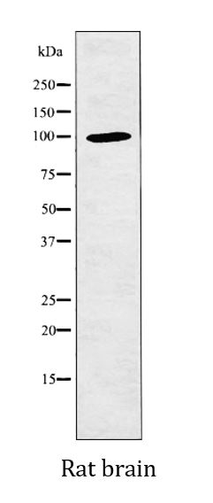
| Positive Control | Rat brain | ||||
| Observed Size | 100 kDa |
Properties
| Form | Liquid |
|---|---|
| Purification | Affinity purification with phospho-specific peptide and the non-phospho specific antibodies were removed by chromatography using non-phosphopeptide. |
| Buffer | PBS (pH 7.4), 150 mM NaCl, 0.02% Sodium azide and 50% Glycerol. |
| Preservative | 0.02% Sodium azide |
| Stabilizer | 50% Glycerol |
| Concentration | 1 mg/ml |
| Storage Instruction | For continuous use, store undiluted antibody at 2-8°C for up to a week. For long-term storage, aliquot and store at -20°C. Storage in frost free freezers is not recommended. Avoid repeated freeze/thaw cycles. Suggest spin the vial prior to opening. The antibody solution should be gently mixed before use. |
| Note | For laboratory research only, not for drug, diagnostic or other use. |
Bioinformation
| Database Links |
Swiss-port # Q13469 Human Nuclear factor of activated T-cells, cytoplasmic 2 Swiss-port # Q60591 Mouse Nuclear factor of activated T-cells, cytoplasmic 2 |
|---|---|
| Gene Symbol | NFATC2 |
| Gene Full Name | nuclear factor of activated T-cells, cytoplasmic, calcineurin-dependent 2 |
| Background | This gene is a member of the nuclear factor of activated T cells (NFAT) family. The product of this gene is a DNA-binding protein with a REL-homology region (RHR) and an NFAT-homology region (NHR). This protein is present in the cytosol and only translocates to the nucleus upon T cell receptor (TCR) stimulation, where it becomes a member of the nuclear factors of activated T cells transcription complex. This complex plays a central role in inducing gene transcription during the immune response. Alternate transcriptional splice variants encoding different isoforms have been characterized. [provided by RefSeq, Apr 2012] |
| Function | Plays a role in the inducible expression of cytokine genes in T-cells, especially in the induction of the IL-2, IL-3, IL-4, TNF-alpha or GM-CSF. Promotes invasive migration through the activation of GPC6 expression and WNT5A signaling pathway. [UniProt] |
| Cellular Localization | Cytoplasm. Nucleus. Note=Cytoplasmic for the phosphorylated form and nuclear after activation that is controlled by calcineurin-mediated dephosphorylation. Rapid nuclear exit of NFATC is thought to be one mechanism by which cells distinguish between sustained and transient calcium signals. The subcellular localization of NFATC plays a key role in the regulation of gene transcription. [UniProt] |
| Calculated MW | 100 kDa |
| PTM | In resting cells, phosphorylated by NFATC-kinase on at least 18 sites in the 99-363 region. Upon cell stimulation, all these sites except Ser-243 are dephosphorylated by calcineurin. Dephosphorylation induces a conformational change that simultaneously exposes an NLS and masks an NES, which results in nuclear localization. Simultaneously, Ser-53 or Ser-56 is phosphorylated; which is required for full transcriptional activity. Ubiquitinated in endothelial cells by RNF213 downstream of the non-canonical Wnt signaling pathway, leading to its degradation by the proteasome. [UniProt] |
Images (1) Click the Picture to Zoom In
-
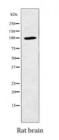
ARG43375 anti-NFATc2 / NFAT1 phospho (Ser326) antibody WB image
Western blot: Rat brain lysate stained with ARG43375 anti-NFATc2 / NFAT1 phospho (Ser326) antibody.