ARG52221
anti-NF66 / alpha Internexin antibody
anti-NF66 / alpha Internexin antibody for ICC/IF,IHC-Frozen sections,Western blot and Human,Mouse,Rat,Cow,Mammal,Pig
Neuroscience antibody
Overview
| Product Description | Chicken Polyclonal antibody recognizes NF66 / alpha Internexin |
|---|---|
| Tested Reactivity | Hu, Ms, Rat, Cow, Mamm, Pig |
| Tested Application | ICC/IF, IHC-Fr, WB |
| Host | Chicken |
| Clonality | Polyclonal |
| Isotype | IgY |
| Target Name | NF66 / alpha Internexin |
| Antigen Species | Rat |
| Immunogen | Recombinant rat alpha-internexin expressed in and purified from E. coli |
| Conjugation | Un-conjugated |
| Alternate Names | Neurofilament 5; Neurofilament-66; Alpha-Inx; NEF5; NF-66; Alpha-internexin; 66 kDa neurofilament protein; TXBP-1 |
Application Instructions
| Application Suggestion |
|
||||||||
|---|---|---|---|---|---|---|---|---|---|
| Application Note | Specific for the ~66k alpha Internexin protein. Minor bands at ~150k are probably covalent dimers and bands at ~50k represent alpha-internexin breakdown products. * The dilutions indicate recommended starting dilutions and the optimal dilutions or concentrations should be determined by the scientist. |
||||||||
| Observed Size | ~ 62 kDa |
Properties
| Form | Liquid |
|---|---|
| Purification | Total IgY fraction |
| Buffer | Total IgY fraction in PBS and 10 mM Sodium azide |
| Preservative | 10 mM Sodium azide |
| Storage Instruction | For continuous use, store undiluted antibody at 2-8°C for up to a week. For long-term storage, aliquot and store at -20°C or below. Storage in frost free freezers is not recommended. Avoid repeated freeze/thaw cycles. Suggest spin the vial prior to opening. The antibody solution should be gently mixed before use. |
| Note | For laboratory research only, not for drug, diagnostic or other use. |
Bioinformation
| Database Links | |
|---|---|
| Gene Symbol | INA |
| Gene Full Name | internexin neuronal intermediate filament protein, alpha |
| Background | Alpha-internexin is a Class IV intermediate filament originally discovered as it co-purifies with other neurofilament subunits. Alpha-internexin is related to but distinct from the better known neurofilament triplet proteins, NF-L, NF-M and NF-H, having similar protein sequence motifs and a similar intron organization. It is expressed only in neurons and in large amounts early in neuronal development, but is down-regulated in many neurons as development proceeds. Many classes of mature neurons contain alpha-internexin in addition to NF-L, NF-M and NF-H. In some mature neurons alphainternexin is the only neurofilament subunit expressed. Antibodies to alpha-internexin are therefore unique probes to study and classify neuronal types and follow their processes in sections and in tissue culture. In addition, recent studies show a marked up-regulation of alpha-internexin during neuronal regeneration. The use of antibodies to this protein in the study of brain tumors has not been examined to date, but is likely to be of interest. Recently Cairns et al. used this antibody to show that alphainternexin is an abundant component of the inclusions of neurofilament inclusion body disease (NFID), a serious human neurodegenerative disorder. The antibody was also used to confirm the presence of circulating auto-antibodies to alpha-internexin in the sera of some patients with endocrine autoimmunity, as well as in some normal individuals. |
| Research Area | Neuroscience antibody |
| Calculated MW | 55 kDa |
| PTM | O-glycosylated. |
Images (4) Click the Picture to Zoom In
-
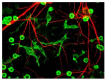
ARG52221 anti-NF66 / alpha Internexin antibody ICC/IF image
Immunofluorescence: Cultured neurons and glia showing specific labeling of neuronal processes (red) using ARG52221 anti-NF66 / alpha Internexin antibody and microglia (green) with a coronin 1a antibody.
-
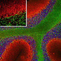
ARG52221 anti-NF66 / alpha Internexin antibody IHC-Fr image
Immunohistochemistry: Frozen section of Rat cerebellum tissue stained with ARG52221 anti-NF66 / alpha Internexin antibody (red) at 1:5000 dilution, and co-stained with anti-MBP antibody (green) at 1:5000 dilution. Hoechst (blue) for nuclear staining. (Sample preparation: Following transcardial perfusion of Rat with 4% paraformaldehyde, brain was post fixed for 24 hours, cut to 45 µM, and free-floating sections were stained with the above antibodies.)
The alpha Internexin antibody selectively stains axons and dendrites of neuronal cells, in particular Purkinje cells, parallel fibers and the axons of granule cells, while the MBP antibody stains myelin sheathes around axons.
-
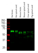
ARG52221 anti-NF66 / alpha Internexin antibody WB image
Western blot: Rat brain, Rat spinal cord, Mouse brain, Mouse spinal cord, Cow spinal cord and Pig spinal cord lysates stained with ARG52221 anti-NF66 / alpha Internexin antibody (green) at 1:10000 dilution.
-
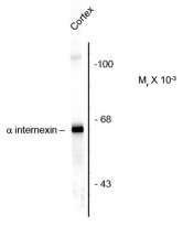
ARG52221 anti-NF66 / alpha Internexin antibody WB image
Western blot: Rat cortex lysate showing specific immunolabeling of the ~66 kDa alpha internexin protein stained with ARG52221 anti-NF66 / alpha Internexin antibody.


