ARG64265
anti-NCF4 / P40PHOX antibody
anti-NCF4 / P40PHOX antibody for IHC-Formalin-fixed paraffin-embedded sections,Western blot and Human
Cell Biology and Cellular Response antibody; Metabolism antibody; Signaling Transduction antibody
Overview
| Product Description | Goat Polyclonal antibody recognizes NCF4 / P40PHOX |
|---|---|
| Tested Reactivity | Hu |
| Tested Application | IHC-P, WB |
| Specificity | This antibody is expected to recognise both reported isoforms (NP_000622.2 and NP_038202.1). |
| Host | Goat |
| Clonality | Polyclonal |
| Isotype | IgG |
| Target Name | NCF4 / P40PHOX |
| Antigen Species | Human |
| Immunogen | C-KDFPEEDDPTN |
| Conjugation | Un-conjugated |
| Alternate Names | p40phox; SH3PXD4; Neutrophil NADPH oxidase factor 4; Neutrophil cytosol factor 4; P40PHOX; SH3 and PX domain-containing protein 4; p40-phox; NCF; NCF-4 |
Application Instructions
| Application Suggestion |
|
||||||
|---|---|---|---|---|---|---|---|
| Application Note | WB: Recommend incubate at RT for 1h. IHC-P: Antigen Retrieval: Steam tissue section in Citrate buffer (pH 6.0). * The dilutions indicate recommended starting dilutions and the optimal dilutions or concentrations should be determined by the scientist. |
Properties
| Form | Liquid |
|---|---|
| Purification | Purified from goat serum by antigen affinity chromatography. |
| Buffer | Tris saline (pH 7.3), 0.02% Sodium azide and 0.5% BSA. |
| Preservative | 0.02% Sodium azide |
| Stabilizer | 0.5% BSA |
| Concentration | 0.5 mg/ml |
| Storage Instruction | For continuous use, store undiluted antibody at 2-8°C for up to a week. For long-term storage, aliquot and store at -20°C or below. Storage in frost free freezers is not recommended. Avoid repeated freeze/thaw cycles. Suggest spin the vial prior to opening. The antibody solution should be gently mixed before use. |
| Note | For laboratory research only, not for drug, diagnostic or other use. |
Bioinformation
| Database Links | |
|---|---|
| Background | The protein encoded by this gene is a cytosolic regulatory component of the superoxide-producing phagocyte NADPH-oxidase, a multicomponent enzyme system important for host defense. This protein is preferentially expressed in cells of myeloid lineage. It interacts primarily with neutrophil cytosolic factor 2 (NCF2/p67-phox) to form a complex with neutrophil cytosolic factor 1 (NCF1/p47-phox), which further interacts with the small G protein RAC1 and translocates to the membrane upon cell stimulation. This complex then activates flavocytochrome b, the membrane-integrated catalytic core of the enzyme system. The PX domain of this protein can bind phospholipid products of the PI(3) kinase, which suggests its role in PI(3) kinase-mediated signaling events. The phosphorylation of this protein was found to negatively regulate the enzyme activity. Alternatively spliced transcript variants encoding distinct isoforms have been observed. [provided by RefSeq, Jul 2008] |
| Research Area | Cell Biology and Cellular Response antibody; Metabolism antibody; Signaling Transduction antibody |
| Calculated MW | 39 kDa |
Images (4) Click the Picture to Zoom In
-
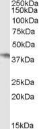
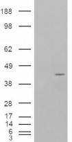
ARG64265 anti-NCF4 / P40PHOX antibody WB image
Western Blot: Daudi cell lysate (35 µg protein in RIPA buffer) stained with ARG64265 anti-NCF4 / P40PHOX antibody at 0.5 µg/ml dilution.
-
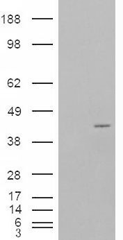
ARG64265 anti-NCF4 / P40PHOX antibody WB image
Western Blot: 1). Mock transfection; 2) P40PHOX (RC201191) expressing plasmid transfected HEK293 cell lysate standed with ARG64265 anti-NCF4 / P40PHOX antibody
-
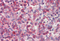
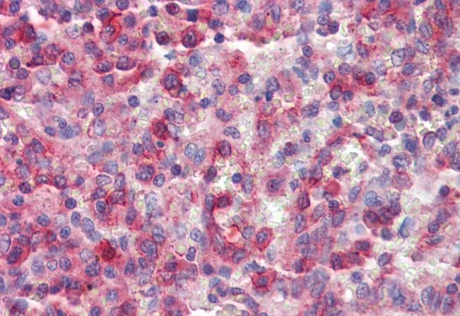
ARG64265 anti-NCF4 / P40PHOX antibody IHC-P image
Immunohistochemistry: Paraffin-embedded Human spleen tissue. Antigen Retrieval: Steam tissue section in Citrate buffer (pH 6.0). The tissue section was stained with ARG64265 anti-NCF4 / P40PHOX antibody at 2.5 µg/ml dilution followed by AP-staining.
-
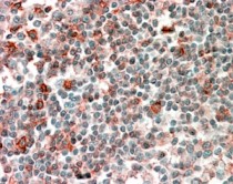
ARG64265 anti-NCF4 / P40PHOX antibody IHC-P image
Immunohistochemistry: paraffin embedded Human Tonsil. (Steamed antigen retrieval with citrate buffer pH 6) stained with ARG64265 anti-NCF4 / P40PHOX antibody at 2.5 µg/ml dilution followed by AP-staining.