ARG63148
anti-NCF1 / p47phox antibody
anti-NCF1 / p47phox antibody for Flow cytometry,ICC/IF,IHC-Formalin-fixed paraffin-embedded sections,Western blot and Human,Mouse,Pig
Cancer antibody; Metabolism antibody; Signaling Transduction antibody
Overview
| Product Description | Goat Polyclonal antibody recognizes NCF1 / p47phox |
|---|---|
| Tested Reactivity | Hu, Ms, Pig |
| Predict Reactivity | Cow, Rat |
| Tested Application | FACS, ICC/IF, IHC-P, WB |
| Host | Goat |
| Clonality | Polyclonal |
| Isotype | IgG |
| Target Name | NCF1 / p47phox |
| Antigen Species | Human |
| Immunogen | C-SESTKRKLASAV |
| Conjugation | Un-conjugated |
| Alternate Names | Nox organizer 2; SH3PXD1A; NCF-47K; 47 kDa autosomal chronic granulomatous disease protein; NOXO2; Nox-organizing protein 2; 47 kDa neutrophil oxidase factor; Neutrophil cytosol factor 1; SH3 and PX domain-containing protein 1A; p47phox; NCF1A; Neutrophil NADPH oxidase factor 1; p47-phox; NCF-1 |
Application Instructions
| Application Suggestion |
|
||||||||||
|---|---|---|---|---|---|---|---|---|---|---|---|
| Application Note | WB: Recommend incubate at RT for 1h. IHC-P: Antigen Retrieval: Steam tissue section in Citrate buffer (pH 6.0). * The dilutions indicate recommended starting dilutions and the optimal dilutions or concentrations should be determined by the scientist. |
Properties
| Form | Liquid |
|---|---|
| Purification | Purified from goat serum by ammonium sulphate precipitation followed by antigen affinity chromatography using the immunizing peptide. |
| Buffer | Tris saline (pH 7.3), 0.02% Sodium azide and 0.5% BSA |
| Preservative | 0.02% Sodium azide |
| Stabilizer | 0.5% BSA |
| Concentration | 0.5 mg/ml |
| Storage Instruction | For continuous use, store undiluted antibody at 2-8°C for up to a week. For long-term storage, aliquot and store at -20°C or below. Storage in frost free freezers is not recommended. Avoid repeated freeze/thaw cycles. Suggest spin the vial prior to opening. The antibody solution should be gently mixed before use. |
| Note | For laboratory research only, not for drug, diagnostic or other use. |
Bioinformation
| Database Links | |
|---|---|
| Background | The protein encoded by this gene is a 47 kDa cytosolic subunit of neutrophil NADPH oxidase. This oxidase is a multicomponent enzyme that is activated to produce superoxide anion. Mutations in this gene have been associated with chronic granulomatous disease. [provided by RefSeq, Jul 2008] |
| Research Area | Cancer antibody; Metabolism antibody; Signaling Transduction antibody |
| Calculated MW | 45 kDa |
| PTM | Phosphorylated by PRKCD; phosphorylation induces activation of NCF1 and NADPH oxidase activity. |
Images (6) Click the Picture to Zoom In
-
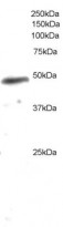
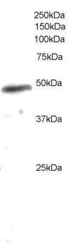
ARG63148 anti-NCF1 / p47phox antibody WB image
Western Blot: U937 lysate (35 µg protein in RIPA buffer) stained with ARG63148 anti-NCF1 / p47phox antibody at 0.2 µg/ml dilution.
-
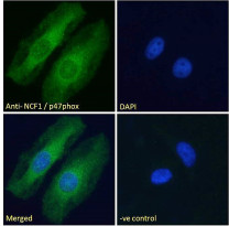
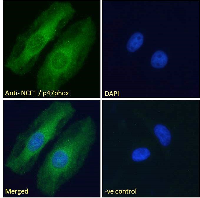
ARG63148 anti-NCF1 / p47phox antibody ICC/IF image
Immunofluorescence: Paraformaldehyde fixed HeLa cells permeabilized with 0.15% Triton. Cells were stained with ARG63148 anti-NCF1 / p47phox antibody (green) at 10 µg/ml dilution for 1 hour. DAPI (blue) for nuclear staining. Negative control: Unimmunized goat IgG (green) at 10 µg/ml dilution.
-

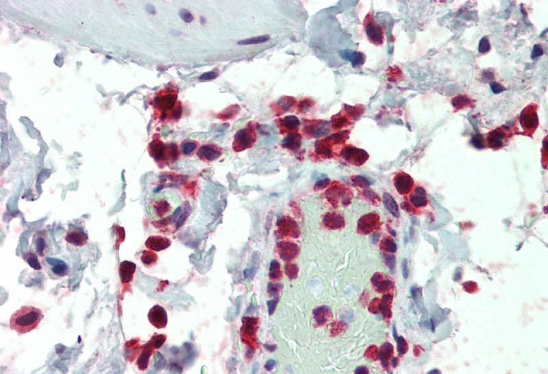
ARG63148 anti-NCF1 / p47phox antibody IHC-P image
Immunohistochemistry: Paraffin-embedded Human colon tissue. Antigen Retrieval: Steam tissue section in Citrate buffer (pH 6.0). The tissue section was stained with ARG63148 anti-NCF1 / p47phox antibody at 5 µg/ml dilution followed by AP-staining.
-
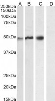
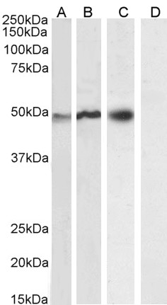
ARG63148 anti-NCF1 / p47phox antibody WB image
Western blot: 35 µg of U937 (A), Daudi (B), U251 (C) and A431 (D, negative control) cell lysates (in RIPA buffer) stained with ARG63148 anti-NCF1 / p47phox antibody at 0.2 µg/ml (A), 0.01 µg/ml (B) and 0.3 µg/ml (C, D) dilutions and incubated at RT for 1 hour.
-
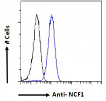
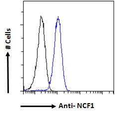
ARG63148 anti-NCF1 / p47phox antibody FACS image
Flow Cytometry: Paraformaldehyde-fixed HeLa cells permeabilized with 0.5% Triton. Cells were stained with ARG63148 anti-NCF1 / p47phox antibody (blue line) at 10 µg/ml dilution for 1 hour, followed by incubation with Alexa FluorR 488 labelled secondary antibody. IgG control: Unimmunized goat IgG (black line).
-
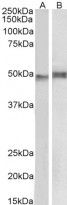
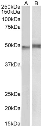
ARG63148 anti-NCF1 / p47phox antibody WB image
Western blot: 35 µg of Mouse thymus (A) and Pig spleen (B) lysates (in RIPA buffer) stained with ARG63148 anti-NCF1 / p47phox antibody at 1 µg/ml dilution and incubated at RT for 1 hour.