ARG58568
anti-NALP3 / Cryopyrin antibody
anti-NALP3 / Cryopyrin antibody for Flow cytometry,ICC/IF,IHC-Formalin-fixed paraffin-embedded sections,Western blot and Human,Rat
NLRP3 Inflammasome Study antibody
Overview
| Product Description | Rabbit Polyclonal antibody recognizes NALP3 / Cryopyrin |
|---|---|
| Tested Reactivity | Hu, Rat |
| Predict Reactivity | Ms, Hm |
| Tested Application | FACS, ICC/IF, IHC-P, WB |
| Host | Rabbit |
| Clonality | Polyclonal |
| Isotype | IgG |
| Target Name | NALP3 / Cryopyrin |
| Antigen Species | Human |
| Immunogen | Synthetic peptide corresponding to aa. 12-31 of Human NALP3 / Cryopyrin. (RYLEDLEDVDLKKFKMHLED) |
| Conjugation | Un-conjugated |
| Alternate Names | MWS; FCAS; CLR1.1; CIAS1; FCU; AGTAVPRL; C1orf7; Caterpiller protein 1.1; PYRIN-containing APAF1-like protein 1; AVP; Angiotensin/vasopressin receptor AII/AVP-like; Cold autoinflammatory syndrome 1 protein; NALP3; AII; FCAS1; NACHT, LRR and PYD domains-containing protein 3; Cryopyrin; PYPAF1 |
Application Instructions
| Application Suggestion |
|
||||||||||
|---|---|---|---|---|---|---|---|---|---|---|---|
| Application Note | IHC-P: Antigen Retrieval: Heat mediation was performed in Citrate buffer (pH 6.0) for 20 min. * The dilutions indicate recommended starting dilutions and the optimal dilutions or concentrations should be determined by the scientist. |
||||||||||
| Observed Size | ~ 118 kDa |
Properties
| Form | Liquid |
|---|---|
| Purification | Affinity purification with immunogen. |
| Buffer | 0.9% NaCl, 0.2% Na2HPO4, 0.05% Thimerosal, 0.05% Sodium azide and 5% BSA. |
| Preservative | 0.05% Thimerosal and 0.05% Sodium azide |
| Stabilizer | 5% BSA |
| Concentration | 0.5 mg/ml |
| Storage Instruction | For continuous use, store undiluted antibody at 2-8°C for up to a week. For long-term storage, aliquot and store at -20°C or below. Storage in frost free freezers is not recommended. Avoid repeated freeze/thaw cycles. Suggest spin the vial prior to opening. The antibody solution should be gently mixed before use. |
| Note | For laboratory research only, not for drug, diagnostic or other use. |
Bioinformation
| Database Links |
Swiss-port # Q96P20 Human NACHT, LRR and PYD domains-containing protein 3 |
|---|---|
| Gene Symbol | NLRP3 |
| Gene Full Name | NLR family, pyrin domain containing 3 |
| Background | NALP3 gene encodes a pyrin-like protein containing a pyrin domain, a nucleotide-binding site (NBS) domain, and a leucine-rich repeat (LRR) motif. This protein interacts with the apoptosis-associated speck-like protein PYCARD/ASC, which contains a caspase recruitment domain, and is a member of the NALP3 inflammasome complex. This complex functions as an upstream activator of NF-kappaB signaling, and it plays a role in the regulation of inflammation, the immune response, and apoptosis. Mutations in this gene are associated with familial cold autoinflammatory syndrome (FCAS), Muckle-Wells syndrome (MWS), chronic infantile neurological cutaneous and articular (CINCA) syndrome, and neonatal-onset multisystem inflammatory disease (NOMID). Multiple alternatively spliced transcript variants encoding distinct isoforms have been identified for this gene. Alternative 5' UTR structures are suggested by available data; however, insufficient evidence is available to determine if all of the represented 5' UTR splice patterns are biologically valid. [provided by RefSeq, Oct 2008] |
| Function | NALP3: As the sensor component of the NLRP3 inflammasome, plays a crucial role in innate immunity and inflammation. In response to pathogens and other damage-associated signals, initiates the formation of the inflammasome polymeric complex, made of NLRP3, PYCARD and CASP1 (and possibly CASP4 and CASP5). Recruitment of proCASP1 to the inflammasome promotes its activation and CASP1-catalyzed IL1B and IL18 maturation and secretion in the extracellular milieu (PubMed:28847925). Activation of NLRP3 inflammasome is also required for HMGB1 secretion (PubMed:22801494). The active cytokines and HMGB1 stimulate inflammatory responses. Inflammasomes can also induce pyroptosis, an inflammatory form of programmed cell death. Under resting conditions, NLRP3 is autoinhibited. NLRP3 activation stimuli include extracellular ATP, reactive oxygen species, K(+) efflux, crystals of monosodium urate or cholesterol, amyloid-beta fibers, environmental or industrial particles and nanoparticles, cytosolic dsRNA, etc. However, it is unclear what constitutes the direct NLRP3 activator. Activation in presence of cytosolic dsRNA is mediated by DHX33 (PubMed:23871209). Independently of inflammasome activation, regulates the differentiation of T helper 2 (Th2) cells and has a role in Th2 cell-dependent asthma and tumor growth. During Th2 differentiation, required for optimal IRF4 binding to IL4 promoter and for IRF4-dependent IL4 transcription. Binds to the consensus DNA sequence 5'-GRRGGNRGAG-3'. May also participate in the transcription of IL5, IL13, GATA3, CCR3, CCR4 and MAF. [UniProt] |
| Cellular Localization | Cytoplasm. [UniProt] |
| Highlight | Related products: NALP3 antibodies; NALP3 ELISA Kits; NALP3 Duos / Panels; Anti-Rabbit IgG secondary antibodies; Related news: Exploring Antiviral Immune Response RIP1 activation and pathogenesis of NASH |
| Research Area | NLRP3 Inflammasome Study antibody |
| Calculated MW | 118 kDa |
| PTM | The disulfide bond in the pyrin domain might play a role in reactive oxygen species-mediated activation. Ubiquitinated; undergoes both 'Lys-48'- and 'Lys-63'-linked polyubiquitination. Ubiquitination does not lead to degradation, but inhibits inflammasome activation (By similarity). Deubiquitination is catalyzed by BRCC3 and associated with NLRP3 activation and inflammasome assembly. This process can be induced by the activation of Toll-like receptors (by LPS), through a non-transcriptional pathway dependent on the mitochondrial production of reactive oxygen species, and by ATP. [UniProt] |
Images (6) Click the Picture to Zoom In
-
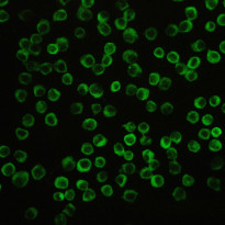
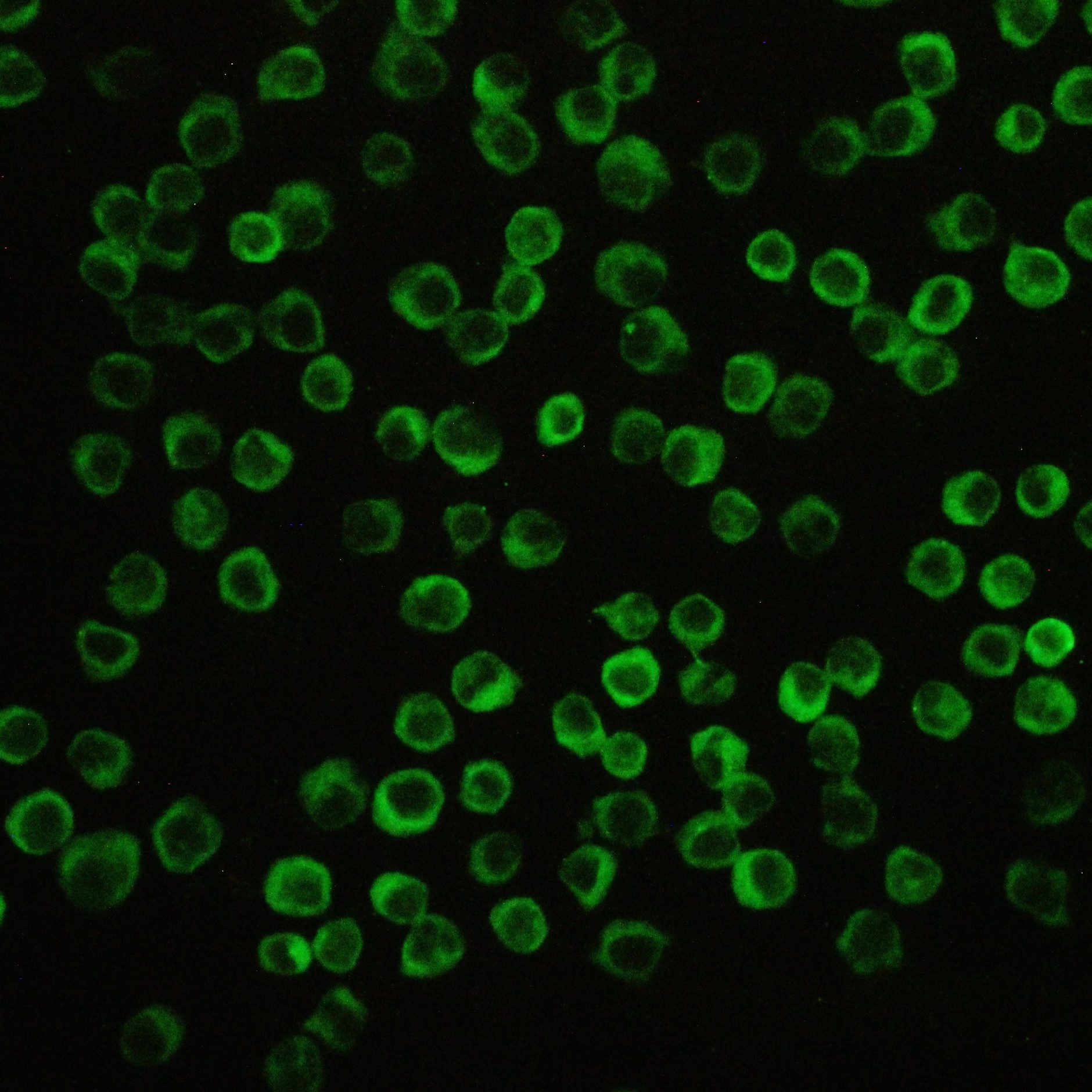
ARG58568 anti-NALP3 / Cryopyrin antibody ICC/IF image
Immunofluorescence: THP-1 cells stained with ARG58568 anti-NALP3 / Cryopyrin antibody (green) at 2 µg/ml, overnight at 4°C.
-
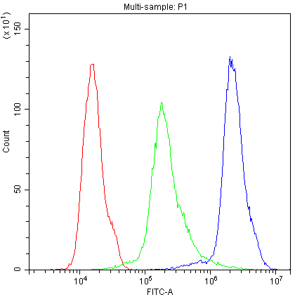
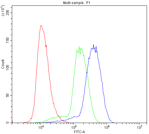
ARG58568 anti-NALP3 / Cryopyrin antibody FACS image
Flow Cytometry: THP-1 cells were blocked with 10% normal Goat serum and stained with ARG58568 anti-NALP3 / Cryopyrin antibody (blue) at 1 µg/10^6 cells for 30 min at 20°C, followed by DyLight®488 labelled secondary antibody. Isotype control antibody (green) was Rabbit IgG at 1 µg/10^6 cells used under the same conditions. Unlabelled sample (red) was also used as a control.
-
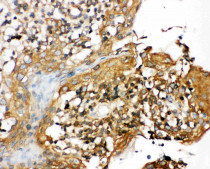
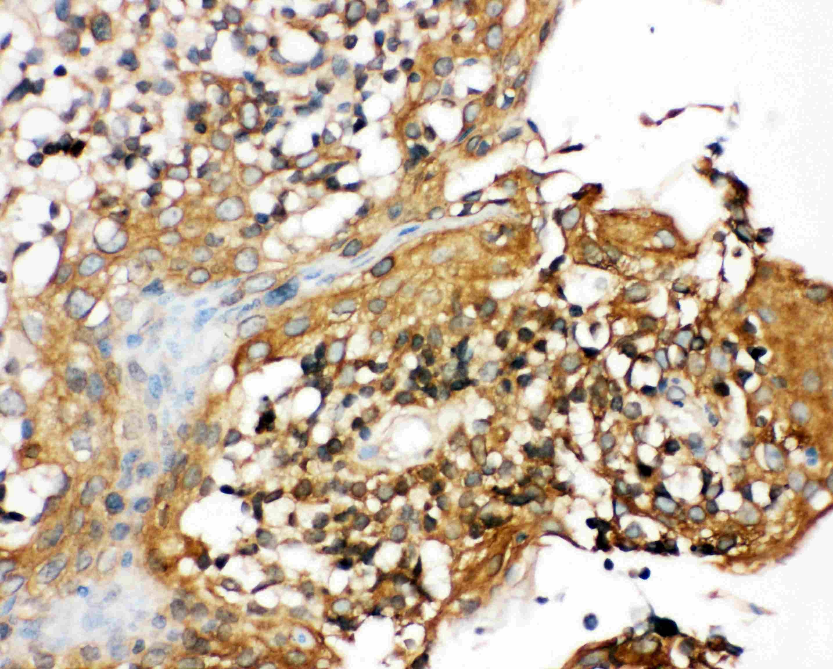
ARG58568 anti-NALP3 / Cryopyrin antibody IHC-P image
Immunohistochemistry: Paraffin-embedded Human tonsil tissue section was blocked with 10% Goat serum. The tissue section was then stained with ARG58568 anti-NALP3 / Cryopyrin antibody at 1 µg/ml, overnight at 4°C. Antigen Retrieval: Heat mediated was performed in Citrate buffer (pH 6.0) for 20 min.
-

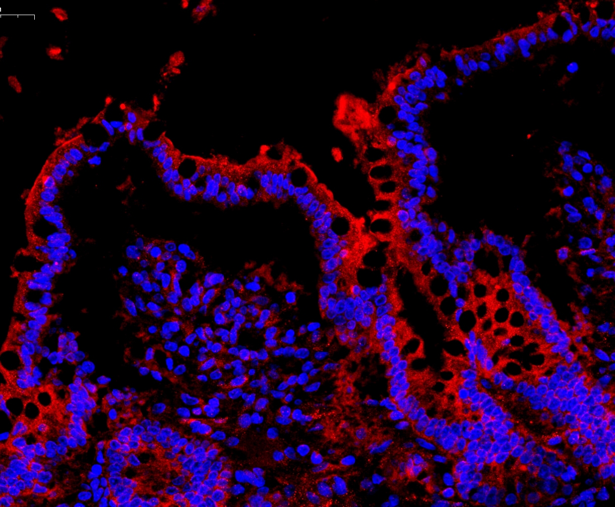
ARG58568 anti-NALP3 / Cryopyrin antibody IHC-P image
Immunohistochemistry: Paraffin-embedded Rat colon tissue. Antigen Retrieval: Heat mediation was performed in Citrate buffer (pH 6.0, epitope retrieval solution) for 20 min. The tissue section was blocked with 10% goat serum. The tissue section was then stained with ARG58568 anti-NALP3 / Cryopyrin antibody (orange-red) at 1μg/ml, overnight at 4°C. The section was counterstained with DAPI (blue).
-
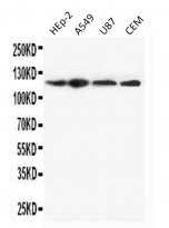
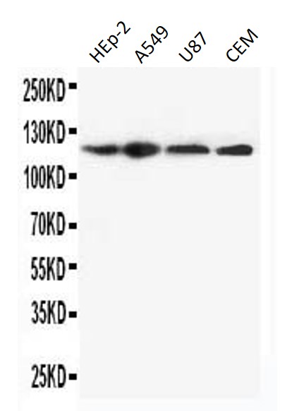
ARG58568 anti-NALP3 / Cryopyrin antibody WB image
Western blot: 50 µg of HEp-2, A549, U87 and CEM cell lysates stained with ARG58568 anti-NALP3 / Cryopyrin antibody at 0.5 µg/ml, overnight at 4°C, under reducing conditions.
-
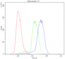
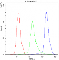
ARG58568 anti-NALP3 / Cryopyrin antibody FACS image
Flow Cytometry: U937 cells were blocked with 10% normal Goat serum and stained with ARG58568 anti-NALP3 / Cryopyrin antibody (blue) at 1 µg/10^6 cells for 30 min at 20°C, followed by DyLight®488 labelled secondary antibody. Isotype control antibody (green) was Rabbit IgG at 1 µg/10^6 cells used under the same conditions. Unlabelled sample (red) was also used as a control.