ARG66189
anti-N Cadherin antibody
anti-N Cadherin antibody for IHC-Formalin-fixed paraffin-embedded sections,Western blot and Human
EMT Study antibody; Mesenchymal Markers antibody
Overview
| Product Description | Rabbit Polyclonal antibody recognizes N Cadherin |
|---|---|
| Tested Reactivity | Hu |
| Predict Reactivity | Ms, Rat |
| Tested Application | IHC-P, WB |
| Specificity | The antibody detects endogenous N-cadherin protein. |
| Host | Rabbit |
| Clonality | Polyclonal |
| Isotype | IgG |
| Target Name | N Cadherin |
| Antigen Species | Human |
| Immunogen | Synthetic peptide around aa. 690-770 of Human N Cadherin. |
| Conjugation | Un-conjugated |
| Alternate Names | Neural cadherin; N-cadherin; CDw325; CDHN; CD antigen CD325; NCAD; Cadherin-2; CD325 |
Application Instructions
| Application Suggestion |
|
||||||
|---|---|---|---|---|---|---|---|
| Application Note | IHC-P: Antigen Retrieval: Boil tissue section in Sodium citrate buffer (pH 6.0) for 20 min. * The dilutions indicate recommended starting dilutions and the optimal dilutions or concentrations should be determined by the scientist. |
Properties
| Form | Liquid |
|---|---|
| Purification | Affinity purification with immunogen. |
| Buffer | PBS, 0.02% Sodium azide, 50% Glycerol and 0.5% BSA. |
| Preservative | 0.02% Sodium azide |
| Stabilizer | 50% Glycerol and 0.5% BSA |
| Concentration | 1 mg/ml |
| Storage Instruction | For continuous use, store undiluted antibody at 2-8°C for up to a week. For long-term storage, aliquot and store at -20°C. Storage in frost free freezers is not recommended. Avoid repeated freeze/thaw cycles. Suggest spin the vial prior to opening. The antibody solution should be gently mixed before use. |
| Note | For laboratory research only, not for drug, diagnostic or other use. |
Bioinformation
| Database Links | |
|---|---|
| Gene Symbol | CDH2 |
| Gene Full Name | cadherin 2, type 1, N-cadherin (neuronal) |
| Background | N Cadherin is a classical cadherin and member of the cadherin superfamily. Alternative splicing results in multiple transcript variants, at least one of which encodes a preproprotein is proteolytically processed to generate a calcium-dependent cell adhesion molecule and glycoprotein. This protein plays a role in the establishment of left-right asymmetry, development of the nervous system and the formation of cartilage and bone. [provided by RefSeq, Nov 2015] |
| Function | N Cadherin is a calcium-dependent cell adhesion protein; preferentially mediates homotypic cell-cell adhesion by dimerization with a CDH2 chain from another cell. Cadherins may thus contribute to the sorting of heterogeneous cell types. Acts as a regulator of neural stem cells quiescence by mediating anchorage of neural stem cells to ependymocytes in the adult subependymal zone: upon cleavage by MMP24, CDH2-mediated anchorage is affected, leading to modulate neural stem cell quiescence. CDH2 may be involved in neuronal recognition mechanism. In hippocampal neurons, may regulate dendritic spine density. [UniProt] |
| Research Area | EMT Study antibody; Mesenchymal Markers antibody |
| Calculated MW | 100 kDa |
| PTM | Cleaved by MMP24. Ectodomain cleavage leads to the generation of a soluble 90 kDa amino-terminal soluble fragment and a 45 kDa membrane-bound carboxy-terminal fragment 1 (CTF1), which is further cleaved by gamma-secretase into a 35 kDa. Cleavage in neural stem cells by MMP24 affects CDH2-mediated anchorage of neural stem cells to ependymocytes in the adult subependymal zone, leading to modulate neural stem cell quiescence (By similarity). May be phosphorylated by OBSCN. |
Images (5) Click the Picture to Zoom In
-

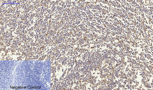
ARG66189 anti-N Cadherin antibody IHC-P image
Immunohistochemistry: Paraffin-embedded Human Tonsil tissue stained with ARG66189 anti-N Cadherin antibody at 1:200 dilution (4°C, overnight). Antigen Retrieval: Boil tissue section in Sodium citrate buffer (pH 6.0) for 20 min.
Negative control was used by secondary antibody only.
-
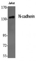
ARG66189 anti-N Cadherin antibody WB image
Western blot: Jurkat cell lysate stained with ARG66189 anti-N Cadherin antibody at 1:1000 dilution.
-
ARG66189 anti-N Cadherin antibody IHC-P image
Immunohistochemistry: Paraffin-embedded Human liver tissue stained with ARG66189 anti-N Cadherin antibody at 1:200 dilution (4°C, overnight). Antigen Retrieval: Boil tissue section in Sodium citrate buffer (pH 6.0) for 20 min.
Negative control was used by secondary antibody only.
-
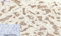
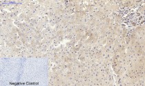
ARG66189 anti-N Cadherin antibody IHC-P image
Immunohistochemistry: Paraffin-embedded Human liver cancer tissue stained with ARG66189 anti-N Cadherin antibody at 1:200 dilution (4°C, overnight). Antigen Retrieval: Boil tissue section in Sodium citrate buffer (pH 6.0) for 20 min.
Negative control was used by secondary antibody only.
-
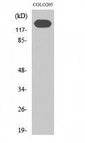
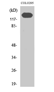
ARG66189 anti-N Cadherin antibody WB image
Western blot: COLO205 cell lysate stained with ARG66189 anti-N Cadherin antibody at 1:1000 dilution.