ARG22587
anti-N Cadherin antibody [13A9]
anti-N Cadherin antibody [13A9] for ICC/IF,IHC-Frozen sections,IHC-Formalin-fixed paraffin-embedded sections,Immunoprecipitation,Western blot and Human,Mouse,Rat
EMT Study antibody; Mesenchymal Markers antibody

Overview
| Product Description | Mouse Monoclonal antibody [13A9] recognizes Cadherin-N/N-Cadherin This antibody recognizes neural cadherin, otherwise known as CD325, a calcium dependent cell-cell adhesion glycoprotein, and member of the cadherin superfamily, which links to the actin cytoskeleton via catenins, and plays a role in cell-matrix adhesion, cell growth and differentiation, and the establishment of left-right asymmetry.N-cadherin is expressed by neurons, endothelial cells, muscle cells, and stem cells, and is one of the primary cadherins recruited to the site of neuronal synapse formation. N-cadherin is directly involved in the differentiation of early hematopoietic progenitors, and is commonly expressed by cancer cells, playing a role in transendothelial migration and metastasis, through the up-regulation of the src kinase pathway, and subsequent failure of the intercellular connection between two adjacent endothelial cells. Mouse anti Human N-cadherin antibody, clone 13A9 studies have demonstrated that expression levels of E-Cadherin and N-Cadherin have a role to play in the invasive properties of breast cancer. Decreased levels of E-cadherin and loss of E-cadherin-mediated adhesion, can result in the transition of a benign epithelial tumor to an invasive tumor, and increase invasiveness, whilst the expression of N-cadherin correlates with motility, invasiveness and tumor metastasis, irrespective of the presence of E-cadherin (Nieman et al. 1999).Mouse anti Human N-cadherin antibody, clone 13A9 has been shown to be specific for N-cadherin, and does not recognize E-cadherin, M-cadherin or P-cadherin (Knudsen et al. 1995). Immunohistological studies have shown that clone 13A9 can be used as a reliable marker for the differential diagnosis of pleural mesotheliomas and lung adenocarcinomas, when used in conjunction with E-cadherin (Han et al. 1997). |
|---|---|
| Tested Reactivity | Hu, Ms, Rat |
| Tested Application | ICC/IF, IHC-Fr, IHC-P, IP, WB |
| Host | Mouse |
| Clonality | Monoclonal |
| Clone | 13A9 |
| Isotype | IgG1 |
| Target Name | N Cadherin |
| Antigen Species | Human |
| Immunogen | Recombinant MBP fusion protein containing the entire cytoplasmic domain of human N-cadherin. |
| Conjugation | Un-conjugated |
| Alternate Names | Neural cadherin; N-cadherin; CD antigen CD325; Cadherin-2 |
Application Instructions
| Application Suggestion |
|
||||||||||||
|---|---|---|---|---|---|---|---|---|---|---|---|---|---|
| Application Note | IHC-P: This product requires antigen retrieval using steam heat treatment prior to staining of paraffin sections. Sodium citrate buffer pH 6.0 is recommended for this purpose. Western Blotting: MCA5698 detects a band of approximately 135-140kDa in human HT-1080 and HeLa cell lysates. * The dilutions indicate recommended starting dilutions and the optimal dilutions or concentrations should be determined by the scientist. |
Properties
| Form | Liquid |
|---|---|
| Purification | Purification with Protein G. |
| Buffer | PBS and 0.09% Sodium azide |
| Preservative | 0.09% Sodium azide |
| Concentration | 1 mg/ml |
| Storage Instruction | For continuous use, store undiluted antibody at 2-8°C for up to a week. For long-term storage, aliquot and store at -20°C or below. Storage in frost free freezers is not recommended. Avoid repeated freeze/thaw cycles. Suggest spin the vial prior to opening. The antibody solution should be gently mixed before use. |
| Note | For laboratory research only, not for drug, diagnostic or other use. |
Bioinformation
| Database Links | |
|---|---|
| Gene Symbol | Cdh2 |
| Gene Full Name | cadherin 2 |
| Background | N Cadherin is a classical cadherin and member of the cadherin superfamily. Alternative splicing results in multiple transcript variants, at least one of which encodes a preproprotein is proteolytically processed to generate a calcium-dependent cell adhesion molecule and glycoprotein. This protein plays a role in the establishment of left-right asymmetry, development of the nervous system and the formation of cartilage and bone. [provided by RefSeq, Nov 2015] |
| Function | N Cadherin is a calcium-dependent cell adhesion protein; preferentially mediates homotypic cell-cell adhesion by dimerization with a CDH2 chain from another cell. Cadherins may thus contribute to the sorting of heterogeneous cell types. Acts as a regulator of neural stem cells quiescence by mediating anchorage of neural stem cells to ependymocytes in the adult subependymal zone: upon cleavage by MMP24, CDH2-mediated anchorage is affected, leading to modulate neural stem cell quiescence. CDH2 may be involved in neuronal recognition mechanism. In hippocampal neurons, may regulate dendritic spine density. [UniProt] |
| Research Area | EMT Study antibody; Mesenchymal Markers antibody |
| Calculated MW | 100 kDa |
| PTM | Cleaved by MMP24. Ectodomain cleavage leads to the generation of a soluble 90 kDa amino-terminal soluble fragment and a 45 kDa membrane-bound carboxy-terminal fragment 1 (CTF1), which is further cleaved by gamma-secretase into a 35 kDa. Cleavage in neural stem cells by MMP24 affects CDH2-mediated anchorage of neural stem cells to ependymocytes in the adult subependymal zone, leading to modulate neural stem cell quiescence (By similarity). May be phosphorylated by OBSCN. |
Images (3) Click the Picture to Zoom In
-
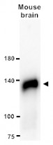
ARG22587 anti-N Cadherin antibody [13A9] WB image
Western blot: 20 µg of Mouse brain lysate stained with ARG22587 anti-N Cadherin antibody [13A9] at 1:1000 dilution.
-
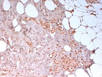
ARG22587 anti-Cadherin-N/N-Cadherin antibody [13A9] IHC-P image
Immunohistochemistry: Formalin-fixed and paraffin-embedded Human breast cancer biopsy stained with ARG22587 anti-Cadherin-N/N-Cadherin antibody [13A9] followed by HRP polymer detection and DAB substrate development. Antigen Retrieval: Boil tissue section in citrate buffer (pH 6.2). (low power).
-
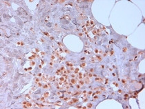
ARG22587 anti-Cadherin-N/N-Cadherin antibody [13A9] IHC-P image
Immunohistochemistry: Formalin-fixed and paraffin-embedded Human breast cancer biopsy stained with ARG22587 anti-Cadherin-N/N-Cadherin antibody [13A9] followed by HRP polymer detection and DAB substrate development. Antigen Retrieval: Boil tissue section in citrate buffer (pH 6.2). (high power).


Customer's Feedback
Excellent

Review for anti-N Cadherin antibody [13A9]
Application:WB
Sample:DLD-1, HCT 116 and HT 29
Sample Loading Amount:20 μg
Primary Antibody Dilution Factor:1:1000
Primary Antibody Incubation Time:overnight
Primary Antibody Incubation Temperature:4 ºC
Conjugation of Secondary Antibody:HRP

Excellent

Review for anti-N Cadherin antibody [13A9]
Application:WB
Sample:Mouse brain
Sample Loading Amount:20 µg
Primary Antibody Dilution Factor:1:1000
Primary Antibody Incubation Time:overnight
Primary Antibody Incubation Temperature:4 ºC
Specific References
Formosanin C suppresses cancer cell proliferation and migration by impeding autophagy machinery
WB / Human


Anticancer Effects and Molecular Mechanisms of Apigenin in Cervical Cancer Cells
WB, IHC-P / Human


