ARG41275
anti-MyD88 antibody
anti-MyD88 antibody for Flow cytometry,IHC-Formalin-fixed paraffin-embedded sections,Western blot and Human,Mouse,Rat
Overview
| Product Description | Rabbit Polyclonal antibody recognizes MyD88 |
|---|---|
| Tested Reactivity | Hu, Ms, Rat |
| Tested Application | FACS, IHC-P, WB |
| Host | Rabbit |
| Clonality | Polyclonal |
| Isotype | IgG |
| Target Name | MyD88 |
| Antigen Species | Human |
| Immunogen | Recombinant protein corresponding to A44-F264 of Human MyD88. |
| Conjugation | Un-conjugated |
| Alternate Names | MYD88D; Myeloid differentiation primary response protein MyD88 |
Application Instructions
| Application Suggestion |
|
||||||||
|---|---|---|---|---|---|---|---|---|---|
| Application Note | IHC-P: Antigen Retrieval: Heat mediation was performed in Citrate buffer (pH 6.0, epitope retrieval solution) for 20 min. * The dilutions indicate recommended starting dilutions and the optimal dilutions or concentrations should be determined by the scientist. |
||||||||
| Observed Size | 35 kDa |
Properties
| Form | Liquid |
|---|---|
| Purification | Affinity purification with immunogen. |
| Buffer | 0.2% Na2HPO4, 0.9% NaCl, 0.05% Sodium azide and 5% BSA. |
| Preservative | 0.05% Sodium azide |
| Stabilizer | 5% BSA |
| Concentration | 0.5 mg/ml |
| Storage Instruction | For continuous use, store undiluted antibody at 2-8°C for up to a week. For long-term storage, aliquot and store at -20°C or below. Storage in frost free freezers is not recommended. Avoid repeated freeze/thaw cycles. Suggest spin the vial prior to opening. The antibody solution should be gently mixed before use. |
| Note | For laboratory research only, not for drug, diagnostic or other use. |
Bioinformation
| Database Links | |
|---|---|
| Gene Symbol | MYD88 |
| Gene Full Name | myeloid differentiation primary response 88 |
| Background | This gene encodes a cytosolic adapter protein that plays a central role in the innate and adaptive immune response. This protein functions as an essential signal transducer in the interleukin-1 and Toll-like receptor signaling pathways. These pathways regulate that activation of numerous proinflammatory genes. The encoded protein consists of an N-terminal death domain and a C-terminal Toll-interleukin1 receptor domain. Patients with defects in this gene have an increased susceptibility to pyogenic bacterial infections. Alternate splicing results in multiple transcript variants. [provided by RefSeq, Feb 2010] |
| Function | Adapter protein involved in the Toll-like receptor and IL-1 receptor signaling pathway in the innate immune response. Acts via IRAK1, IRAK2, IRF7 and TRAF6, leading to NF-kappa-B activation, cytokine secretion and the inflammatory response. Increases IL-8 transcription. Involved in IL-18-mediated signaling pathway. Activates IRF1 resulting in its rapid migration into the nucleus to mediate an efficient induction of IFN-beta, NOS2/INOS, and IL12A genes. MyD88-mediated signaling in intestinal epithelial cells is crucial for maintenance of gut homeostasis and controls the expression of the antimicrobial lectin REG3G in the small intestine. [UniProt] |
| Cellular Localization | Cytoplasm. Nucleus. [UniProt] |
| Calculated MW | 33 kDa |
Images (5) Click the Picture to Zoom In
-

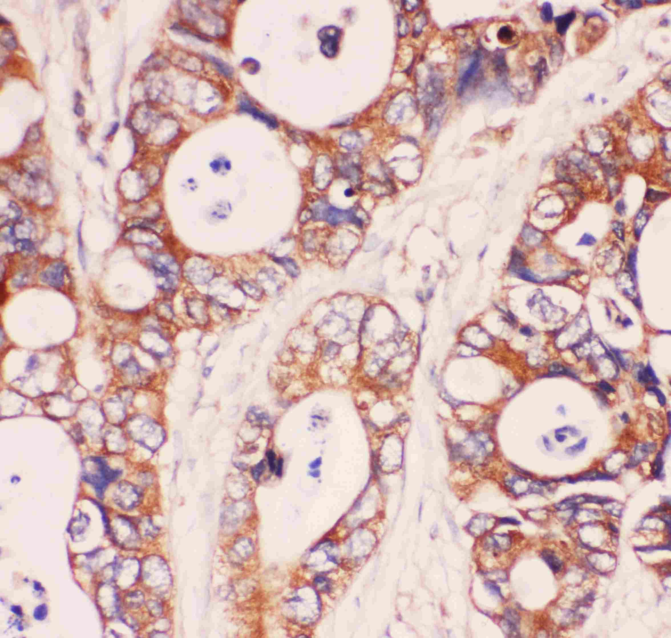
ARG41275 anti-MyD88 antibody IHC-P image
Immunohistochemistry: Paraffin-embedded Human intestinal cancer tissue. Antigen Retrieval: Heat mediation was performed in Citrate buffer (pH 6.0, epitope retrieval solution) for 20 min. The tissue section was blocked with 10% goat serum. The tissue section was then stained with ARG41275 anti-MyD88 antibody at 1 µg/ml dilution, overnight at 4°C.
-
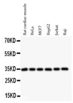
ARG41275 anti-MyD88 antibody WB image
Western blot: 50 µg of samples under reducing conditions. Rat cardiac muscle, HeLa, MCF7, HepG2, Jurkat and Raji whole cell lysates stained with ARG41275 anti-MyD88 antibody at 0.5 µg/ml dilution, overnight at 4°C.
-
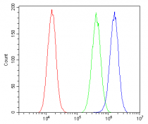
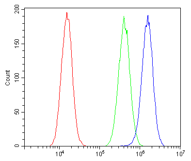
ARG41275 anti-MyD88 antibody FACS image
Flow Cytometry: A549 cells were blocked with 10% normal goat serum and then stained with ARG41275 anti-MyD88 antibody (blue) at 1 µg/10^6 cells for 30 min at 20°C, followed by incubation with DyLight®488 labelled secondary antibody. Isotype control antibody (green) was rabbit IgG (1 µg/10^6 cells) used under the same conditions. Unlabelled sample (red) was also used as a control.
-
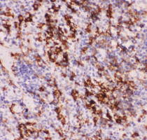
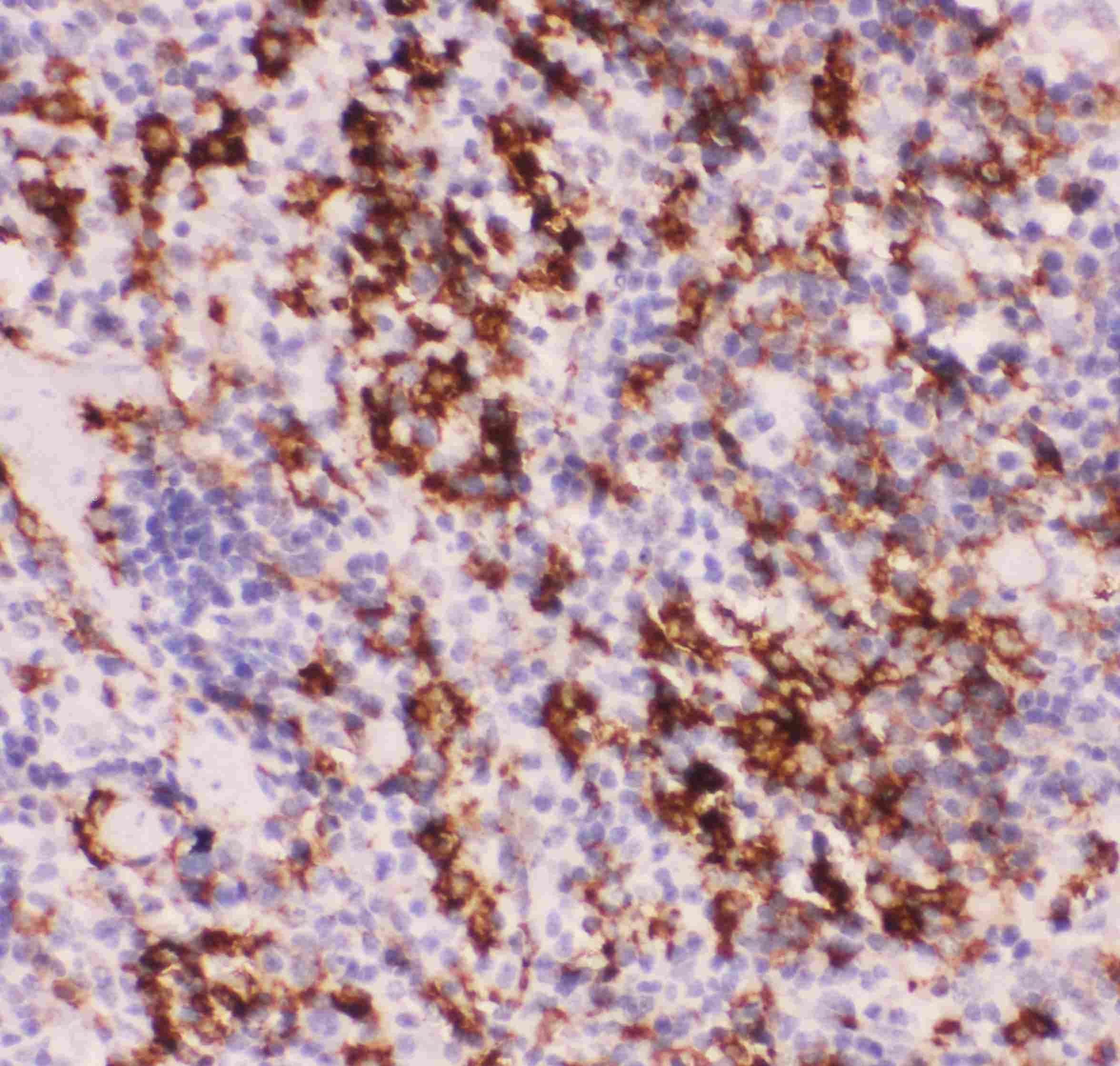
ARG41275 anti-MyD88 antibody IHC-P image
Immunohistochemistry: Paraffin-embedded Mouse spleen tissue. Antigen Retrieval: Heat mediation was performed in Citrate buffer (pH 6.0, epitope retrieval solution) for 20 min. The tissue section was blocked with 10% goat serum. The tissue section was then stained with ARG41275 anti-MyD88 antibody at 1 µg/ml dilution, overnight at 4°C.
-
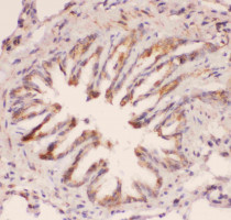
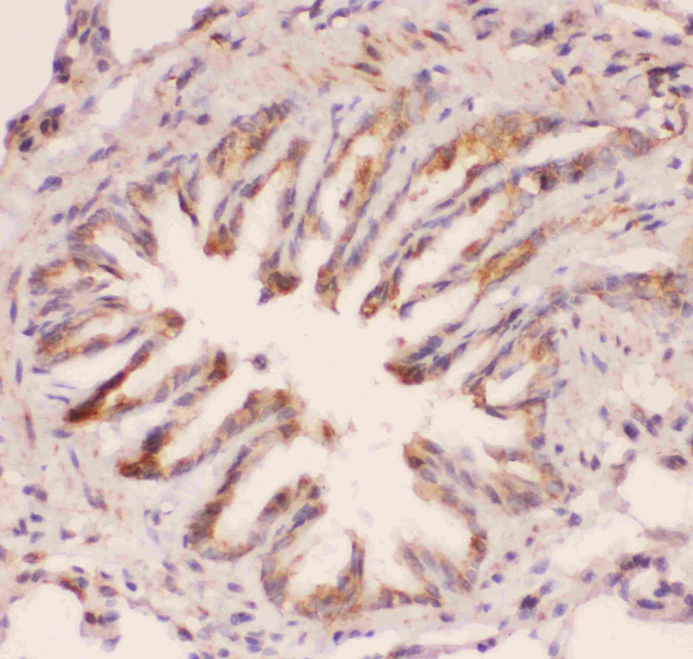
ARG41275 anti-MyD88 antibody IHC-P image
Immunohistochemistry: Paraffin-embedded Rat lung tissue. Antigen Retrieval: Heat mediation was performed in Citrate buffer (pH 6.0, epitope retrieval solution) for 20 min. The tissue section was blocked with 10% goat serum. The tissue section was then stained with ARG41275 anti-MyD88 antibody at 1 µg/ml dilution, overnight at 4°C.

Specific References
Myd88 Signaling Is Involved in the Inflammatory Response in LPS-Induced Mouse Epididymitis and Bone-Marrow-Derived Dendritic Cells
WB / Mouse


