ARG65457
anti-Muscle Actin antibody [HHF35]
anti-Muscle Actin antibody [HHF35] for IHC-Frozen sections,IHC-Formalin-fixed paraffin-embedded sections,Western blot and Human,Mouse,Rat,Cat,Chicken,Dog,Primates,Rabbit
Cancer antibody; Cell Death antibody; Controls and Markers antibody; Signaling Transduction antibody
Overview
| Product Description | Mouse Monoclonal antibody [HHF35] recognizes Muscle Actin |
|---|---|
| Tested Reactivity | Hu, Ms, Rat, Cat, Chk, Dog, NHuPrm, Rb |
| Tested Application | IHC-Fr, IHC-P, WB |
| Specificity | The mouse monoclonal antibody HHF35 recognizes muscle specific alpha and gamma actin (42 kDa) in various species. This antibody stains skeletal, smooth and myocardial cells as well as myoepithelial cells and pericytes of small vessels. It is a widely used marker of muscle and musclederived cells. |
| Host | Mouse |
| Clonality | Monoclonal |
| Clone | HHF35 |
| Isotype | IgG1 |
| Target Name | Muscle Actin |
| Antigen Species | Human |
| Immunogen | SDS extracted protein fraction of Human myocardium. |
| Conjugation | Un-conjugated |
| Alternate Names | CFTDM; MPFD; CFTD; ASMA; NEM1; NEM2; NEM3; Alpha-actin-1; ACTA; CFTD1; Actin, alpha skeletal muscle |
Application Instructions
| Application Suggestion |
|
||||||||
|---|---|---|---|---|---|---|---|---|---|
| Application Note | WB: Under reducing condition. IHC-P: Antigen retrieval steps generally not required, but in case of arterial smooth muscle cells or myoepithelial cells, pepsin or trypsin pretreatment is recommended. * The dilutions indicate recommended starting dilutions and the optimal dilutions or concentrations should be determined by the scientist. |
||||||||
| Positive Control | WB: Positive control: Murine femoral muscle and heart. Negative control: HUVEC. |
Properties
| Form | Liquid |
|---|---|
| Purification | Purified from cell culture supernatant by protein-A affinity chromatography. |
| Purity | > 95% (by SDS-PAGE) |
| Buffer | PBS (pH 7.4) and 15 mM Sodium azide |
| Preservative | 15 mM Sodium azide |
| Concentration | 1 mg/ml |
| Storage Instruction | For continuous use, store undiluted antibody at 2-8°C for up to a week. For long-term storage, aliquot and store at -20°C or below. Storage in frost free freezers is not recommended. Avoid repeated freeze/thaw cycles. Suggest spin the vial prior to opening. The antibody solution should be gently mixed before use. |
| Note | For laboratory research only, not for drug, diagnostic or other use. |
Bioinformation
| Database Links | |
|---|---|
| Gene Symbol | ACTA1 |
| Gene Full Name | actin, alpha 1, skeletal muscle |
| Background | Actin is a highly conserved ubiquitous globular protein (G-actin) that polymerizes to form fibrous F-actin microfilaments. In higher eucaryotes several actin isoforms have been identified, that fall into three classes. Alpha actin is a structural component of the contractile apparatus of muscle cells or muscle-derived cells. Beta actin and gamma actin play roles in regulation of cell motility in other cell types. Specific subcellular structures such as as stress fibers, focal adhesions, filopodia etc., are formed by involvement of actin cytoskeleton. |
| Function | Actins are highly conserved proteins that are involved in various types of cell motility and are ubiquitously expressed in all eukaryotic cells. [UniProt] |
| Research Area | Cancer antibody; Cell Death antibody; Controls and Markers antibody; Signaling Transduction antibody |
| Calculated MW | 42 kDa |
| PTM | Oxidation of Met-46 and Met-49 by MICALs (MICAL1, MICAL2 or MICAL3) to form methionine sulfoxide promotes actin filament depolymerization. MICAL1 and MICAL2 produce the (R)-S-oxide form. The (R)-S-oxide form is reverted by MSRB1 and MSRB2, which promote actin repolymerization (By similarity). Monomethylation at Lys-86 (K84me1) regulates actin-myosin interaction and actomyosin-dependent processes. Demethylation by ALKBH4 is required for maintaining actomyosin dynamics supporting normal cleavage furrow ingression during cytokinesis and cell migration. (Microbial infection) Monomeric actin is cross-linked by V.cholerae toxins RtxA and VgrG1 in case of infection: bacterial toxins mediate the cross-link between Lys-52 of one monomer and Glu-272 of another actin monomer, resulting in formation of highly toxic actin oligomers that cause cell rounding (PubMed:19015515). The toxin can be highly efficient at very low concentrations by acting on formin homology family proteins: toxic actin oligomers bind with high affinity to formins and adversely affect both nucleation and elongation abilities of formins, causing their potent inhibition in both profilin-dependent and independent manners (PubMed:26228148). |
Images (2) Click the Picture to Zoom In
-
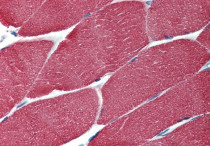
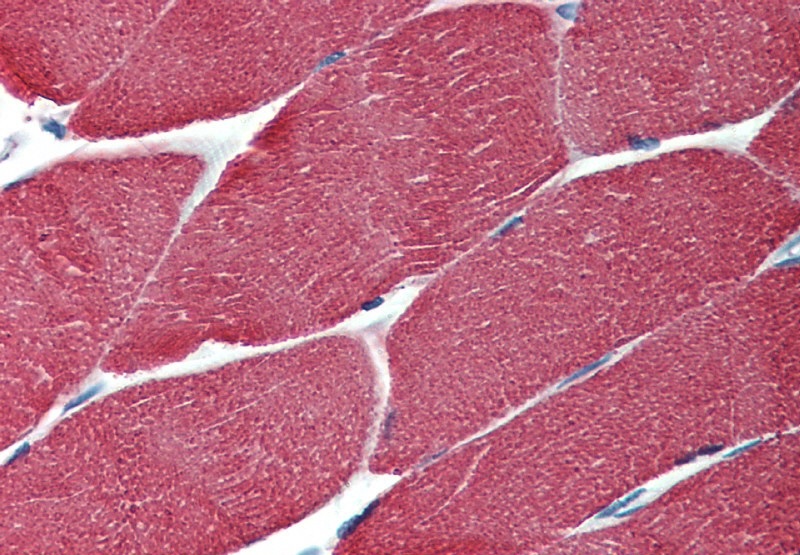
ARG65457 anti-Muscle Actin antibody [HHF35] IHC-P image
Immunohistochemistry: Paraffin-embedded Human muscle tissue stained with ARG65457 anti-Muscle Actin antibody [HHF35].
-
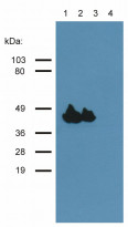
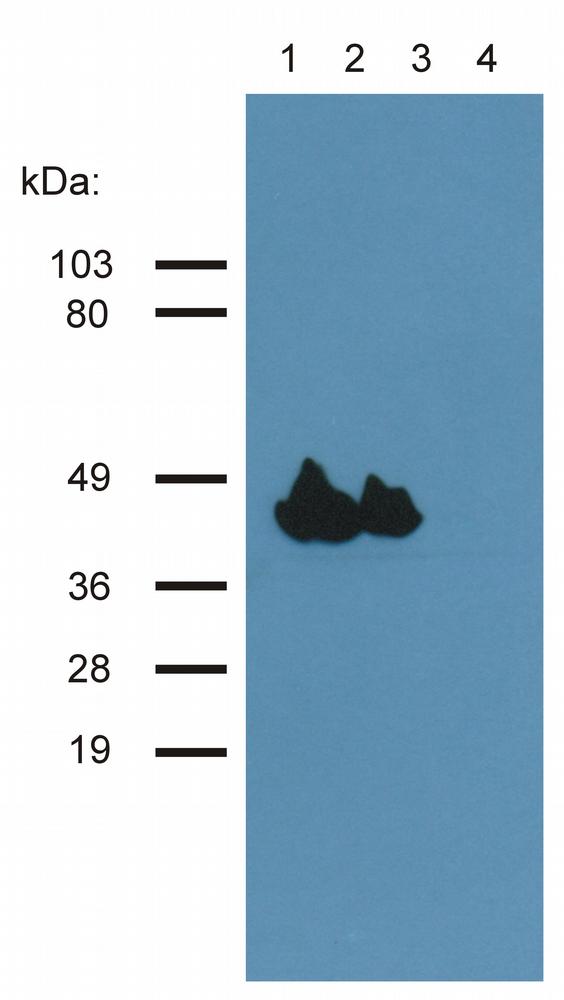
ARG65457 anti-Muscle Actin antibody [HHF35] WB image
Western blot: 1) Murine femoral muscle, 2) Murine heart, 3) HeLa cell lysate, and 4) HUVEC cell lysate stained with ARG65457 anti-Muscle Actin antibody [HHF35].
Clone References
Live cell imaging reveals actin-cytoskeleton-induced self-association of the actin-bundling protein WLIM1.
WB / Human


Expression of E-cadherin in angiomyolipoma.
IHC / Human
Fascin expression and its potential significance in gastrointestinal stromal tumors.
IHC / Human
G-CSF promotes the proliferation of developing cardiomyocytes in vivo and in derivation from ESCs and iPSCs.
ICC/IF / Mouse


