ARG52341
anti-Mitochondria antibody [113-1]
anti-Mitochondria antibody [113-1] for Flow cytometry,ICC/IF,IHC-Formalin-fixed paraffin-embedded sections,Western blot and Human
Controls and Markers antibody; Metabolism antibody; Signaling Transduction antibody
Overview
| Product Description | Mouse Monoclonal antibody [113-1] recognizes Mitochondria |
|---|---|
| Tested Reactivity | Hu |
| Species Does Not React With | Ms, Rat |
| Tested Application | FACS, ICC/IF, IHC-P, WB |
| Host | Mouse |
| Clonality | Monoclonal |
| Clone | 113-1 |
| Isotype | IgG1 |
| Target Name | Mitochondria |
| Antigen Species | Human |
| Immunogen | Human cell homogenate |
| Conjugation | Un-conjugated |
Application Instructions
| Application Suggestion |
|
||||||||||
|---|---|---|---|---|---|---|---|---|---|---|---|
| Application Note | Specific for human mitochondria in all cell types. The antibody works on acetone fixed and 2% paraformaldehyde fixed cells. The antibody also works for immunohistochemistry on 2% paraformaldehyde fixed (5-10 minutes) cryostat sections. * The dilutions indicate recommended starting dilutions and the optimal dilutions or concentrations should be determined by the scientist. |
Properties
| Form | Liquid |
|---|---|
| Purification | Total IgG fraction |
| Buffer | Total IgG fraction |
| Storage Instruction | For continuous use, store undiluted antibody at 2-8°C for up to a week. For long-term storage, aliquot and store at -20°C or below. Storage in frost free freezers is not recommended. Avoid repeated freeze/thaw cycles. Suggest spin the vial prior to opening. The antibody solution should be gently mixed before use. |
| Note | For laboratory research only, not for drug, diagnostic or other use. |
Bioinformation
| Background | Mitochondria are most commonly known as the power plants of the cell as they produce ATP, but they are also involved in many other important cellular processes such as cell signaling, growth and differentiation (McBride et al., 2006). In addition, mitochondria have been shown to play a role in apoptosis (Green 1998). This antibody is an excellent marker for human cells in xenographic model research. It reacts specifically with human cells, including neurons and embryonic stem cells. |
|---|---|
| Research Area | Controls and Markers antibody; Metabolism antibody; Signaling Transduction antibody |
Images (1) Click the Picture to Zoom In
-
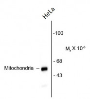
ARG52341 anti-Mitochondria antibody [113-1] WB image
Western blot: HeLa cell lysate showing specific immunolabeling of the ~60 kDa mitochondrial protein stained with ARG52341 anti-Mitochondria antibody [113-1].

Clone References
Overexpression of the cell adhesion molecule claudin-9 is associated with invasion in pituitary oncocytomas.


Identification of a subtype-specific ENC1 gene related to invasiveness in human pituitary null cell adenoma and oncocytomas.
A tissue-engineered humanized xenograft model of human breast cancer metastasis to bone.
IHC-P / Human
Characterization of mesenchymal stem cells from human normal and hyperplastic gingiva.
IHC-P / Human


