ARG43408
anti-Midkine antibody
anti-Midkine antibody for ICC/IF,IHC-Formalin-fixed paraffin-embedded sections,Immunoprecipitation,Western blot and Human
Overview
| Product Description | Rabbit Polyclonal antibody recognizes Midkine |
|---|---|
| Tested Reactivity | Hu |
| Tested Application | ICC/IF, IHC-P, IP, WB |
| Host | Rabbit |
| Clonality | Polyclonal |
| Isotype | IgG |
| Target Name | Midkine |
| Antigen Species | Human |
| Immunogen | Synthetic peptide derived from Human Midkine. |
| Conjugation | Un-conjugated |
| Alternate Names | ARAP; Neurite outgrowth-promoting protein; Midkine; MK; Neurite outgrowth-promoting factor 2; Amphiregulin-associated protein; Midgestation and kidney protein; NEGF2 |
Application Instructions
| Application Suggestion |
|
||||||||||
|---|---|---|---|---|---|---|---|---|---|---|---|
| Application Note | * The dilutions indicate recommended starting dilutions and the optimal dilutions or concentrations should be determined by the scientist. | ||||||||||
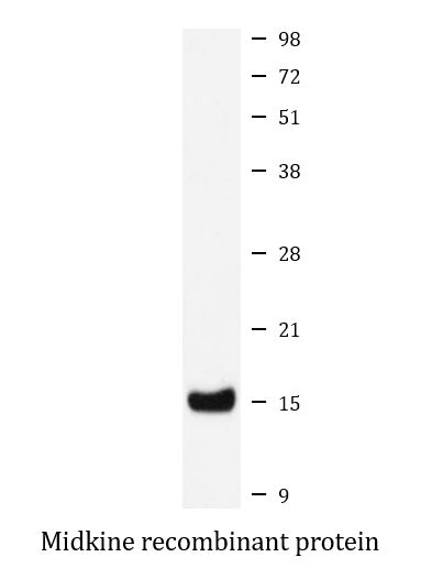
| Observed Size | ~ 15 kDa |
Properties
| Form | Liquid |
|---|---|
| Purification | Affinity purified. |
| Buffer | PBS (pH 7.4), 150 mM NaCl, 0.02% Sodium azide and 50% Glycerol. |
| Preservative | 0.02% Sodium azide |
| Stabilizer | 50% Glycerol |
| Storage Instruction | For continuous use, store undiluted antibody at 2-8°C for up to a week. For long-term storage, aliquot and store at -20°C. Storage in frost free freezers is not recommended. Avoid repeated freeze/thaw cycles. Suggest spin the vial prior to opening. The antibody solution should be gently mixed before use. |
| Note | For laboratory research only, not for drug, diagnostic or other use. |
Bioinformation
| Database Links | |
|---|---|
| Gene Symbol | MDK |
| Gene Full Name | midkine (neurite growth-promoting factor 2) |
| Background | This gene encodes a member of a small family of secreted growth factors that binds heparin and responds to retinoic acid. The encoded protein promotes cell growth, migration, and angiogenesis, in particular during tumorigenesis. This gene has been targeted as a therapeutic for a variety of different disorders. Alternatively spliced transcript variants encoding multiple isoforms have been observed. [provided by RefSeq, Jul 2012] |
| Function | Secreted protein that functions as cytokine and growth factor and mediates its signal through cell-surface proteoglycan and non-proteoglycan receptors (PubMed:18469519, PubMed:12573468, PubMed:12122009, PubMed:10212223, PubMed:24458438, PubMed:15466886, PubMed:12084985, PubMed:10772929). Binds cell-surface proteoglycan receptors via their chondroitin sulfate (CS) groups (PubMed:12084985, PubMed:10212223). Thereby regulates many processes like inflammatory response, cell proliferation, cell adhesion, cell growth, cell survival, tissue regeneration, cell differentiation and cell migration (PubMed:12573468, PubMed:12122009, PubMed:10212223, PubMed:10683378, PubMed:24458438, PubMed:22323540, PubMed:12084985, PubMed:15466886, PubMed:10772929). Participates in inflammatory processes by exerting two different activities. Firstly, mediates neutrophils and macrophages recruitment to the sites of inflammation both by direct action by cooperating namely with ITGB2 via LRP1 and by inducing chemokine expression (PubMed:10683378, PubMed:24458438). This inflammation can be accompanied by epithelial cell survival and smooth muscle cell migration after renal and vessel damage, respectively (PubMed:10683378). Secondly, suppresses the development of tolerogenic dendric cells thereby inhibiting the differentiation of regulatory T cells and also promote T cell expansion through NFAT signaling and Th1 cell differentiation (PubMed:22323540). Promotes tissue regeneration after injury or trauma. After heart damage negatively regulates the recruitment of inflammatory cells and mediates cell survival through activation of anti-apoptotic signaling pathways via MAPKs and AKT pathways through the activation of angiogenesis (By similarity). Also facilitates liver regeneration as well as bone repair by recruiting macrophage at trauma site and by promoting cartilage development by facilitating chondrocyte differentiation (By similarity). Plays a role in brain by promoting neural precursor cells survival and growth through interaction with heparan sulfate proteoglycans (By similarity). Binds PTPRZ1 and promotes neuronal migration and embryonic neurons survival (PubMed:10212223). Binds SDC3 or GPC2 and mediates neurite outgrowth and cell adhesion (PubMed:12084985, PubMed:1768439). Binds chondroitin sulfate E and heparin leading to inhibition of neuronal cell adhesion induced by binding with GPC2 (PubMed:12084985). Binds CSPG5 and promotes elongation of oligodendroglial precursor-like cells (By similarity). Also binds ITGA6:ITGB1 complex; this interaction mediates MDK-induced neurite outgrowth (PubMed:15466886, PubMed:1768439). Binds LRP1; promotes neuronal survival (PubMed:10772929). Binds ITGA4:ITGB1 complex; this interaction mediates MDK-induced osteoblast cells migration through PXN phosphorylation (PubMed:15466886). Binds anaplastic lymphoma kinase (ALK) which induces ALK activation and subsequent phosphorylation of the insulin receptor substrate (IRS1), followed by the activation of mitogen-activated protein kinase (MAPK) and PI3-kinase, and the induction of cell proliferation (PubMed:12122009). Promotes epithelial to mesenchymal transition through interaction with NOTCH2 (PubMed:18469519). During arteriogenesis, plays a role in vascular endothelial cell proliferation by inducing VEGFA expression and release which in turn induces nitric oxide synthase expression. Moreover activates vasodilation through nitric oxide synthase activation (By similarity). Negatively regulates bone formation in response to mechanical load by inhibiting Wnt/beta-catenin signaling in osteoblasts (By similarity). In addition plays a role in hippocampal development, working memory, auditory response, early fetal adrenal gland development and the female reproductive system (By similarity). [UniProt] |
| Cellular Localization | Secreted. [UniProt] |
| Calculated MW | 16 kDa |
Images (2) Click the Picture to Zoom In
-
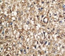
ARG43408 anti-Midkine antibody IHC-P image
Immunohistochemistry: Paraffin-embedded Human liver carcinoma tissue stained with ARG43408 anti-Midkine antibody.
-
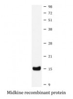
ARG43408 anti-Midkine antibody WB image
Western blot: Midkine recombinant protein stained with ARG43408 anti-Midkine antibody.