ARG66419
anti-Met antibody [SQab18121]
anti-Met antibody [SQab18121] for ELISA,Flow cytometry,ICC/IF,IHC-Formalin-fixed paraffin-embedded sections,Western blot and Human
Overview
| Product Description | Mouse Monoclonal antibody [SQab18121] recognizes Met |
|---|---|
| Tested Reactivity | Hu |
| Tested Application | ELISA, FACS, ICC/IF, IHC-P, WB |
| Host | Mouse |
| Clonality | Monoclonal |
| Clone | SQab18121 |
| Isotype | IgG |
| Target Name | Met |
| Antigen Species | Human |
| Immunogen | Recombinant Human Met protein. |
| Conjugation | Un-conjugated |
| Alternate Names | Scatter factor receptor; c-Met; HGF receptor; HGFR; EC 2.7.10.1; SF receptor; AUTS9; Proto-oncogene c-Met; Tyrosine-protein kinase Met; HGF/SF receptor; Hepatocyte growth factor receptor; RCCP2; DFNB97 |
Application Instructions
| Application Suggestion |
|
||||||||||||
|---|---|---|---|---|---|---|---|---|---|---|---|---|---|
| Application Note | * The dilutions indicate recommended starting dilutions and the optimal dilutions or concentrations should be determined by the scientist. |
Properties
| Purification | Affinity purification with immunogen. |
|---|---|
| Buffer | PBS (pH 7.4) and 0.01% Thimerosal. |
| Preservative | 0.01% Thimerosal |
| Concentration | 1 mg/ml |
| Storage Instruction | For continuous use, store undiluted antibody at 2-8°C for up to a week. For long-term storage, aliquot and store at -20°C or below. Storage in frost free freezers is not recommended. Avoid repeated freeze/thaw cycles. Suggest spin the vial prior to opening. The antibody solution should be gently mixed before use. |
| Note | For laboratory research only, not for drug, diagnostic or other use. |
Bioinformation
| Database Links | |
|---|---|
| Gene Symbol | MET |
| Gene Full Name | MET proto-oncogene, receptor tyrosine kinase |
| Background | The proto-oncogene MET product is the hepatocyte growth factor receptor and encodes tyrosine-kinase activity. The primary single chain precursor protein is post-translationally cleaved to produce the alpha and beta subunits, which are disulfide linked to form the mature receptor. Various mutations in the MET gene are associated with papillary renal carcinoma. Two transcript variants encoding different isoforms have been found for this gene. [provided by RefSeq, Jul 2008] |
| Function | Receptor tyrosine kinase that transduces signals from the extracellular matrix into the cytoplasm by binding to hepatocyte growth factor/HGF ligand. Regulates many physiological processes including proliferation, scattering, morphogenesis and survival. Ligand binding at the cell surface induces autophosphorylation of MET on its intracellular domain that provides docking sites for downstream signaling molecules. Following activation by ligand, interacts with the PI3-kinase subunit PIK3R1, PLCG1, SRC, GRB2, STAT3 or the adapter GAB1. Recruitment of these downstream effectors by MET leads to the activation of several signaling cascades including the RAS-ERK, PI3 kinase-AKT, or PLCgamma-PKC. The RAS-ERK activation is associated with the morphogenetic effects while PI3K/AKT coordinates prosurvival effects. During embryonic development, MET signaling plays a role in gastrulation, development and migration of muscles and neuronal precursors, angiogenesis and kidney formation. In adults, participates in wound healing as well as organ regeneration and tissue remodeling. Promotes also differentiation and proliferation of hematopoietic cells. Acts as a receptor for Listeria internalin inlB, mediating entry of the pathogen into cells. [UniProt] |
| Cellular Localization | Membrane; Single-pass type I membrane protein. Isoform 3: Secreted. [UniProt] |
| Calculated MW | 156 kDa |
| PTM | Autophosphorylated in response to ligand binding on Tyr-1234 and Tyr-1235 in the kinase domain leading to further phosphorylation of Tyr-1349 and Tyr-1356 in the C-terminal multifunctional docking site. Dephosphorylated by PTPRJ at Tyr-1349 and Tyr-1365. Dephosphorylated by PTPN1 and PTPN2. Ubiquitinated. Ubiquitination by CBL regulates MET endocytosis, resulting in decreasing plasma membrane receptor abundance, and in endosomal degradation and/or recycling of internalized receptors. [UniProt] |
Images (4) Click the Picture to Zoom In
-
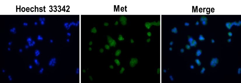
ARG66419 anti-Met antibody [SQab18121] ICC/IF image
Immunofluorescence: MCF7 cells were fixed in 100% methanol, permeabilized with PBS containing 0.1% Triton X-100. Cells were stained with ARG66419 anti-Met antibody [SQab18121] (green) at 1:200 dilution and cell nuclei were stained with Hoechst 33342 (blue).
-
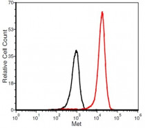
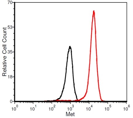
ARG66419 anti-Met antibody [SQab18121] FACS image
Flow Cytometry: MCF-7 cells were stained with ARG66419 anti-Met antibody [SQab18121] at 2 µg/ml dilution (red) and without antibody control (black).
-
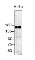
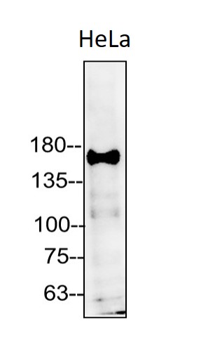
ARG66419 anti-Met antibody [SQab18121] WB image
Western blot: 50 µg of HeLa cell lysate stained with ARG66419 anti-Met antibody [SQab18121] at 1:5000 dilution.
-
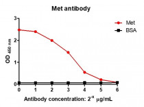
ARG66419 anti-Met antibody [SQab18121] ELISA image
ELISA: Titration curve of ARG66419 anti-Met antibody [SQab18121]. Red: Met; Black: BSA (negative control).