ARG41406
anti-Melanoma gp100 antibody
anti-Melanoma gp100 antibody for IHC-Formalin-fixed paraffin-embedded sections,Western blot and Human
Overview
| Product Description | Rabbit Polyclonal antibody recognizes Melanoma gp100 |
|---|---|
| Tested Reactivity | Hu |
| Tested Application | IHC-P, WB |
| Host | Rabbit |
| Clonality | Polyclonal |
| Isotype | IgG |
| Target Name | Melanoma gp100 |
| Antigen Species | Human |
| Immunogen | Synthetic peptide derived from Human Melanoma gp100. |
| Conjugation | Un-conjugated |
| Alternate Names | Premelanosome protein; SILV; ME20; Melanocyte protein Pmel 17; ME20-M; Secreted melanoma-associated ME20 antigen; 95 kDa melanocyte-specific secreted glycoprotein; Silver locus protein homolog; ME20S; D12S53E; SIL; P1; Melanocyte protein PMEL; PMEL17; ME20-S; Melanoma-associated ME20 antigen; gp100; ME20M; P100; SI; P26; Melanocytes lineage-specific antigen GP100 |
Application Instructions
| Application Suggestion |
|
||||||
|---|---|---|---|---|---|---|---|
| Application Note | * The dilutions indicate recommended starting dilutions and the optimal dilutions or concentrations should be determined by the scientist. | ||||||
| Observed Size | 100 kDa (full length) or 26 kda (C-terminal fragment) |
Properties
| Form | Liquid |
|---|---|
| Purification | Affinity purified. |
| Buffer | PBS (pH 7.4), 150 mM NaCl, 0.02% Sodium azide and 50% Glycerol. |
| Preservative | 0.02% Sodium azide |
| Stabilizer | 50% Glycerol |
| Storage Instruction | For continuous use, store undiluted antibody at 2-8°C for up to a week. For long-term storage, aliquot and store at -20°C. Storage in frost free freezers is not recommended. Avoid repeated freeze/thaw cycles. Suggest spin the vial prior to opening. The antibody solution should be gently mixed before use. |
| Note | For laboratory research only, not for drug, diagnostic or other use. |
Bioinformation
| Database Links | |
|---|---|
| Gene Symbol | PMEL |
| Gene Full Name | premelanosome protein |
| Background | This gene encodes a melanocyte-specific type I transmembrane glycoprotein. The encoded protein is enriched in melanosomes, which are the melanin-producing organelles in melanocytes, and plays an essential role in the structural organization of premelanosomes. This protein is involved in generating internal matrix fibers that define the transition from Stage I to Stage II melanosomes. This protein undergoes a complex pattern of prosttranslational processing and modification that is essential to the proper functioning of the protein. A secreted form of this protein that is released by proteolytic ectodomain shedding may be used as a melanoma-specific serum marker. Alternate splicing results in multiple transcript variants. [provided by RefSeq, Jan 2011] |
| Function | Plays a central role in the biogenesis of melanosomes. Involved in the maturation of melanosomes from stage I to II. The transition from stage I melanosomes to stage II melanosomes involves an elongation of the vesicle, and the appearance within of distinct fibrillar structures. Release of the soluble form, ME20-S, could protect tumor cells from antibody mediated immunity. [UniProt] |
| Cellular Localization | ER membrane; Single-pass type I membrane protein. Golgi apparatus. Melanosome. Endosome, multivesicular body. Note=Identified by mass spectrometry in melanosome fractions from stage I to stage IV. Localizes predominantly to intralumenal vesicles within multivesicular bodies. Associates with ILVs found within the lumen of premelanosomes and melanosomes and particularly in compartments that serve as precursors to the striated stage II premelanosomes. M-alpha: Secreted. [UniProt] |
| Calculated MW | 70 kDa |
| PTM | A small amount of P1/P100 (major form) undergoes glycosylation to yield P2/P120 (minor form). P2 is cleaved by a furin-like proprotein convertase (PC) in a pH-dependent manner in a post-Golgi, prelysosomal compartment into two disulfide-linked subunits: a large lumenal subunit, M-alpha/ME20-S, and an integral membrane subunit, M-beta. Despite cleavage, only a small fraction of M-alpha is secreted, whereas most M-alpha and M-beta remain associated with each other intracellularly. M-alpha is further processed to M-alpha N and M-alpha C. M-alpha C further undergoes processing to yield M-alpha C1 and M-alpha C3 (M-alpha C2 in the case of PMEL17-is or PMEL17-ls). Formation of intralumenal fibrils in the melanosomes requires the formation of M-alpha that becomes incorporated into the fibrils. Stage II melanosomes harbor only Golgi-modified Pmel17 fragments that are derived from M-alpha and that bear sialylated O-linked oligosaccharides. N-glycosylated. O-glycosylated; contains sialic acid. [UniProt] |
Images (2) Click the Picture to Zoom In
-
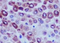
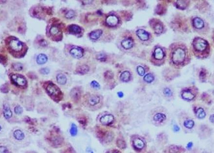
ARG41406 anti-Melanoma gp100 antibody IHC-P image
Immunohistochemistry: Paraffin-embedded Human melanoma tissue stained with ARG41406 anti-Melanoma gp100 antibody.
-
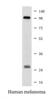
ARG41406 anti-Melanoma gp100 antibody WB image
Western blot: Human melanoma lysate stained with ARG41406 anti-Melanoma gp100 antibody.