ARG59747
anti-MafA antibody
anti-MafA antibody for Western blot and Rat
Overview
| Product Description | Rabbit Polyclonal antibody recognizes MafA |
|---|---|
| Tested Reactivity | Rat |
| Tested Application | WB |
| Host | Rabbit |
| Clonality | Polyclonal |
| Isotype | IgG |
| Target Name | MafA |
| Antigen Species | Human |
| Immunogen | Synthetic peptide corresponding to aa. 136-167 of Human MafA. (EDAVEALIGSGHHGAHHGAHHPAAAAAYEAFR) |
| Conjugation | Un-conjugated |
| Alternate Names | RIPE3b1; Transcription factor MafA; Transcription factor RIPE3b1; V-maf musculoaponeurotic fibrosarcoma oncogene homolog A; hMafA; Pancreatic beta-cell-specific transcriptional activator |
Application Instructions
| Application Suggestion |
|
||||
|---|---|---|---|---|---|
| Application Note | * The dilutions indicate recommended starting dilutions and the optimal dilutions or concentrations should be determined by the scientist. |
Properties
| Form | Liquid |
|---|---|
| Purification | Affinity purification with immunogen. |
| Buffer | 0.9% NaCl, 0.2% Na2HPO4, 0.05% Sodium azide and 5% BSA. |
| Preservative | 0.05% Sodium azide |
| Stabilizer | 5% BSA |
| Concentration | 0.5 mg/ml |
| Storage Instruction | For continuous use, store undiluted antibody at 2-8°C for up to a week. For long-term storage, aliquot and store at -20°C or below. Storage in frost free freezers is not recommended. Avoid repeated freeze/thaw cycles. Suggest spin the vial prior to opening. The antibody solution should be gently mixed before use. |
| Note | For laboratory research only, not for drug, diagnostic or other use. |
Bioinformation
| Gene Symbol | MAFA |
|---|---|
| Gene Full Name | v-maf avian musculoaponeurotic fibrosarcoma oncogene homolog A |
| Background | MAFA is a transcription factor that binds RIPE3b, a conserved enhancer element that regulates pancreatic beta cell-specific expression of the insulin gene (INS; MIM 176730) (Olbrot et al., 2002 [PubMed 12011435]).[supplied by OMIM, Mar 2008] |
| Function | Acts as a transcriptional factor. Specifically binds the insulin enhancer element RIPE3b and activates insulin gene expression. Cooperates synergistically with NEUROD1 and PDX1. Phosphorylation by GSK3 increases its transcriptional activity and is required for its oncogenic activity. Involved either as an oncogene or as a tumor suppressor, depending on the cell context. [UniProt] |
| Cellular Localization | Nucleus. Note=Detected in nuclei of pancreas islet beta cells. [UniProt] |
| Calculated MW | 37 kDa |
| PTM | Ubiquitinated, leading to its degradation by the proteasome. Ser-14 and Ser-65 appear to be the major phosphorylation sites. Phosphorylated by MAPK13 on serine and threonine residues (Probable). Phosphorylation by GSK3 requires prior phosphorylation of Ser-65 by another kinase. Phosphorylation proceeds then from Ser-61 to Thr-57, Thr-53 and Ser-49. GSK3-mediated phosphorylation increases its transcriptional activity through the recruitment of the coactivator PCAF, is required for its transforming activity and leads to its degradation through a ubiquitin/proteasome-dependent pathway. [UniProt] |
Images (1) Click the Picture to Zoom In
-
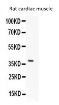
ARG59747 anti-MafA antibody WB image
Western blot: 50 µg of Rat cardiac muscle lysate stained with ARG59747 anti-MafA antibody at 0.5 µg/ml dilution.


