ARG43059
anti-MTA2 antibody
anti-MTA2 antibody for Flow cytometry,ICC/IF,IHC-Formalin-fixed paraffin-embedded sections,Western blot and Human,Mouse,Rat
Overview
| Product Description | Rabbit Polyclonal antibody recognizes MTA2 |
|---|---|
| Tested Reactivity | Hu, Ms, Rat |
| Tested Application | FACS, ICC/IF, IHC-P, WB |
| Host | Rabbit |
| Clonality | Polyclonal |
| Isotype | IgG |
| Target Name | MTA2 |
| Antigen Species | Human |
| Immunogen | Recombinant protein corresponding to M1-D668 of Human MTA2. |
| Conjugation | Un-conjugated |
| Alternate Names | p53 target protein in deacetylase complex; MTA1L1; PID; Metastasis-associated protein MTA2; Metastasis-associated 1-like 1; MTA1-L1 protein |
Application Instructions
| Application Suggestion |
|
||||||||||
|---|---|---|---|---|---|---|---|---|---|---|---|
| Application Note | IHC-P: Antigen Retrieval: Heat mediation was performed in EDTA buffer (pH 8.0). * The dilutions indicate recommended starting dilutions and the optimal dilutions or concentrations should be determined by the scientist. |
Properties
| Form | Liquid |
|---|---|
| Purification | Affinity purification with immunogen. |
| Buffer | 0.2% Na2HPO4, 0.9% NaCl, 0.05% Sodium azide and 4% Trehalose. |
| Preservative | 0.05% Sodium azide |
| Stabilizer | 4% Trehalose |
| Concentration | 0.5 mg/ml |
| Storage Instruction | For continuous use, store undiluted antibody at 2-8°C for up to a week. For long-term storage, aliquot and store at -20°C or below. Storage in frost free freezers is not recommended. Avoid repeated freeze/thaw cycles. Suggest spin the vial prior to opening. The antibody solution should be gently mixed before use. |
| Note | For laboratory research only, not for drug, diagnostic or other use. |
Bioinformation
| Database Links |
Swiss-port # O94776 Human Metastasis-associated protein MTA2 Swiss-port # Q9R190 Mouse Metastasis-associated protein MTA2 |
|---|---|
| Gene Symbol | MTA2 |
| Gene Full Name | metastasis associated 1 family, member 2 |
| Background | This gene encodes a protein that has been identified as a component of NuRD, a nucleosome remodeling deacetylase complex identified in the nucleus of human cells. It shows a very broad expression pattern and is strongly expressed in many tissues. It may represent one member of a small gene family that encode different but related proteins involved either directly or indirectly in transcriptional regulation. Their indirect effects on transcriptional regulation may include chromatin remodeling. It is closely related to another member of this family, a protein that has been correlated with the metastatic potential of certain carcinomas. These two proteins are so closely related that they share the same types of domains. These domains include two DNA binding domains, a dimerization domain, and a domain commonly found in proteins that methylate DNA. One of the proteins known to be a target protein for this gene product is p53. Deacetylation of p53 is correlated with a loss of growth inhibition in transformed cells supporting a connection between these gene family members and metastasis. [provided by RefSeq, May 2011] |
| Function | May be involved in the regulation of gene expression as repressor and activator. The repression might be related to covalent modification of histone proteins. [UniProt] |
| Cellular Localization | Nucleus. [UniProt] |
| Calculated MW | 75 kDa |
Images (5) Click the Picture to Zoom In
-
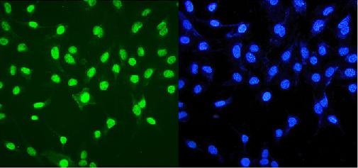
ARG43059 anti-MTA2 antibody ICC/IF image
Immunofluorescence: A549 cells were blocked with 10% goat serum and then stained with ARG43059 anti-MTA2 antibody (green) at 2 µg/ml dilution, overnight at 4°C. DAPI (blue) for nuclear staining.
-
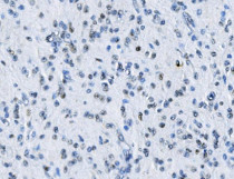
ARG43059 anti-MTA2 antibody IHC-P image
Immunohistochemistry: Paraffin-embedded Human glioma tissue. Antigen Retrieval: Heat mediation was performed in EDTA buffer (pH 8.0). The tissue section was blocked with 10% goat serum. The tissue section was then stained with ARG43059 anti-MTA2 antibody at 1 µg/ml dilution, overnight at 4°C.
-

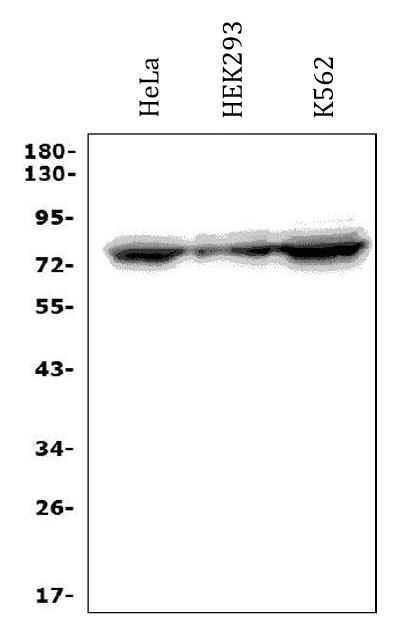
ARG43059 anti-MTA2 antibody WB image
Western blot: 50 µg of sample under reducing conditions. HeLa, HEK293 and K562 whole cell lysates stained with ARG43059 anti-MTA2 antibody at 0.5 µg/ml dilution, overnight at 4°C.
-
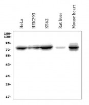
ARG43059 anti-MTA2 antibody WB image
Western blot: 50 µg of sample under reducing conditions. HeLa, HEK293, K562, Rat liver and Mouse heart lysates stained with ARG43059 anti-MTA2 antibody at 0.5 µg/ml dilution, overnight at 4°C.
-
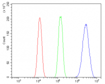
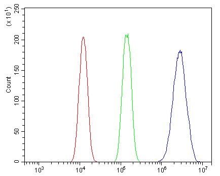
ARG43059 anti-MTA2 antibody FACS image
Flow Cytometry: A431 cells were blocked with 10% normal goat serum and then stained with ARG43059 anti-MTA2 antibody (blue) at 1 µg/10^6 cells for 30 min at 20°C, followed by incubation with DyLight®488 labelled secondary antibody. Isotype control antibody (green) was rabbit IgG (1 µg/10^6 cells) used under the same conditions. Unlabelled sample (red) was also used as a control.