ARG66198
anti-MSH6 antibody [SQab1720]
anti-MSH6 antibody [SQab1720] for Flow cytometry,ICC/IF,IHC-Formalin-fixed paraffin-embedded sections,Immunoprecipitation,Western blot and Human,Rat
DNA Mismatch Repair System Study antibody

Overview
| Product Description | Recombinant Rabbit Monoclonal antibody [SQab1720] recognizes MSH6 |
|---|---|
| Tested Reactivity | Hu, Rat |
| Tested Application | FACS, ICC/IF, IHC-P, IP, WB |
| Host | Rabbit |
| Clonality | Monoclonal |
| Clone | SQab1720 |
| Isotype | IgG |
| Target Name | MSH6 |
| Antigen Species | Human |
| Immunogen | Synthetic peptide around the N-terminus of MSH6. |
| Conjugation | Un-conjugated |
| Alternate Names | DNA mismatch repair protein Msh6; HNPCC5; p160; GTMBP; G/T mismatch-binding protein; HSAP; hMSH6; GTBP; MutS-alpha 160 kDa subunit |
Application Instructions
| Application Suggestion |
|
||||||||||||
|---|---|---|---|---|---|---|---|---|---|---|---|---|---|
| Application Note | IHC-P: Antigen retrieval: Heat mediated was performed using Tris/EDTA buffer pH 9.0. * The dilutions indicate recommended starting dilutions and the optimal dilutions or concentrations should be determined by the scientist. |
Properties
| Form | Liquid |
|---|---|
| Purification | Affinity purification with immunogen. |
| Buffer | PBS, 0.01% Sodium azide, 40% Glycerol and 0.05% BSA. |
| Preservative | 0.01% Sodium azide |
| Stabilizer | 40% Glycerol and 0.05% BSA |
| Storage Instruction | For continuous use, store undiluted antibody at 2-8°C for up to a week. For long-term storage, aliquot and store at -20°C. Storage in frost free freezers is not recommended. Avoid repeated freeze/thaw cycles. Suggest spin the vial prior to opening. The antibody solution should be gently mixed before use. |
| Note | For laboratory research only, not for drug, diagnostic or other use. |
Bioinformation
| Database Links | |
|---|---|
| Gene Symbol | MSH6 |
| Gene Full Name | mutS homolog 6 |
| Background | MSH6 is a member of the DNA mismatch repair MutS family. In E. coli, the MutS protein helps in the recognition of mismatched nucleotides prior to their repair. A highly conserved region of approximately 150 aa, called the Walker-A adenine nucleotide binding motif, exists in MutS homologs. The encoded protein heterodimerizes with MSH2 to form a mismatch recognition complex that functions as a bidirectional molecular switch that exchanges ADP and ATP as DNA mismatches are bound and dissociated. Mutations in this gene may be associated with hereditary nonpolyposis colon cancer, colorectal cancer, and endometrial cancer. Transcripts variants encoding different isoforms have been described. [provided by RefSeq, Jul 2013] |
| Function | MSH6 is a component of the post-replicative DNA mismatch repair system (MMR). Heterodimerizes with MSH2 to form MutS alpha, which binds to DNA mismatches thereby initiating DNA repair. When bound, MutS alpha bends the DNA helix and shields approximately 20 base pairs, and recognizes single base mismatches and dinucleotide insertion-deletion loops (IDL) in the DNA. After mismatch binding, forms a ternary complex with the MutL alpha heterodimer, which is thought to be responsible for directing the downstream MMR events, including strand discrimination, excision, and resynthesis. ATP binding and hydrolysis play a pivotal role in mismatch repair functions. The ATPase activity associated with MutS alpha regulates binding similar to a molecular switch: mismatched DNA provokes ADP-->ATP exchange, resulting in a discernible conformational transition that converts MutS alpha into a sliding clamp capable of hydrolysis-independent diffusion along the DNA backbone. This transition is crucial for mismatch repair. MutS alpha may also play a role in DNA homologous recombination repair. Recruited on chromatin in G1 and early S phase via its PWWP domain that specifically binds trimethylated 'Lys-36' of histone H3 (H3K36me3): early recruitment to chromatin to be replicated allowing a quick identification of mismatch repair to initiate the DNA mismatch repair reaction. [UniProt] |
| Highlight | Related Antibody Duos and Panels: ARG30312 DNA Mismatch Repair System Antibody Duo (MSH2, MSH6) Related products: MSH6 antibodies; MSH6 Duos / Panels; Anti-Rabbit IgG secondary antibodies; Related news: Cancer Pathology Markers (SQ clones) |
| Research Area | DNA Mismatch Repair System Study antibody |
| Calculated MW | 153 kDa |
| PTM | The N-terminus is blocked. Phosphorylated by PRKCZ, which may prevent MutS alpha degradation by the ubiquitin-proteasome pathway. |
Images (3) Click the Picture to Zoom In
-
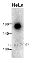
ARG66198 anti-MSH6 antibody [SQab1720] WB image
Western blot: 30 µg of HeLa cell lysate stained with ARG66198 anti-MSH6 antibody [SQab1720] at 1:500 dilution.
-
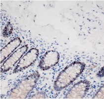
ARG66198 anti-MSH6 antibody [SQab1720] IHC-P image
Immunohistochemistry: Formalin‐fixed and paraffin‐embedded Human colon tissue stained with ARG66198 anti-MSH6 antibody [SQab1720] at 1:500 dilution.
Antigen retrieval: Heat mediated was performed using Tris/EDTA buffer pH 9.0
-
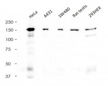
ARG66198 anti-MSH6 antibody [SQab1720] WB image
Western blot: 20 µg of HeLa, A431, SW480, Rat testis and 293HEK cell lysates stained with ARG66198 anti-MSH6 antibody [SQab1720] at 1:5000 dilution.


Customer's Feedback
Excellent

Review for anti-MSH6 antibody [SQab1720]
Application:WB
Sample:HeLa
Sample Loading Amount:30 µg
Primary Antibody Dilution Factor:1:500
Primary Antibody Incubation Time:overnight
Primary Antibody Incubation Temperature:4 ºC


