ARG42832
anti-MR1 antibody
anti-MR1 antibody for Flow cytometry,Western blot and Human
Overview
| Product Description | Rabbit Polyclonal antibody recognizes MR1 |
|---|---|
| Tested Reactivity | Hu |
| Predict Reactivity | Ms, Rat |
| Tested Application | FACS, WB |
| Host | Rabbit |
| Clonality | Polyclonal |
| Isotype | IgG |
| Target Name | MR1 |
| Antigen Species | Human |
| Immunogen | Recombinant protein corresponding to R23-D269 of Human MR1. |
| Conjugation | Un-conjugated |
| Alternate Names | Major histocompatibility complex class I-related gene protein; MHC class I-related gene protein; Class I histocompatibility antigen-like protein; HLALS |
Application Instructions
| Application Suggestion |
|
||||||
|---|---|---|---|---|---|---|---|
| Application Note | * The dilutions indicate recommended starting dilutions and the optimal dilutions or concentrations should be determined by the scientist. | ||||||
| Observed Size | ~ 39 kDa |
Properties
| Form | Liquid |
|---|---|
| Purification | Affinity purification with immunogen. |
| Buffer | 0.2% Na2HPO4, 0.9% NaCl, 0.05% Sodium azide and 4% Trehalose. |
| Preservative | 0.05% Sodium azide |
| Stabilizer | 4% Trehalose |
| Concentration | 0.5 mg/ml |
| Storage Instruction | For continuous use, store undiluted antibody at 2-8°C for up to a week. For long-term storage, aliquot and store at -20°C or below. Storage in frost free freezers is not recommended. Avoid repeated freeze/thaw cycles. Suggest spin the vial prior to opening. The antibody solution should be gently mixed before use. |
| Note | For laboratory research only, not for drug, diagnostic or other use. |
Bioinformation
| Database Links |
Swiss-port # Q95460 Human Major histocompatibility complex class I-related gene protein |
|---|---|
| Gene Symbol | MR1 |
| Gene Full Name | major histocompatibility complex, class I-related |
| Background | MAIT (mucosal-associated invariant T-cells) lymphocytes represent a small population of T-cells primarily found in the gut. The protein encoded by this gene is an antigen-presenting molecule that presents metabolites of microbial vitamin B to MAITs. This presentation may activate the MAITs to regulate the amounts of specific types of bacteria in the gut. Several transcript variants encoding different isoforms have been found for this gene, and a pseudogene of it has been detected about 36 kbp upstream on the same chromosome. [provided by RefSeq, Jul 2015] |
| Function | Antigen-presenting molecule specialized in presenting microbial vitamin B metabolites. Involved in the development and expansion of a small population of T-cells expressing an invariant T-cell receptor alpha chain called mucosal-associated invariant T-cells (MAIT). MAIT lymphocytes are preferentially located in the gut lamina propria and therefore may be involved in monitoring commensal flora or serve as a distress signal. Expression and MAIT cell recognition seem to be ligand-dependent. [UniProt] |
| Cellular Localization | Cell membrane; Single-pass membrane protein; Extracellular side. Endoplasmic reticulum. Isoform 4: Secreted. Isoform 3: Cell membrane; Single-pass type I membrane protein. Endoplasmic reticulum membrane. Note=The larger proportion remains in the ER in an immature state. The subset that reach cell surface does it through a B2M-independent pathway. [UniProt] |
| Calculated MW | 39 kDa |
| PTM | N-glycosylated. [UniProt] |
Images (2) Click the Picture to Zoom In
-
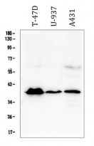
ARG42832 anti-MR1 antibody WB image
Western blot: 50 µg of sample under reducing conditions. T-47D, U-937 and A431 whole cell lysates stained with ARG42832 anti-MR1 antibody at 0.5 µg/ml dilution, overnight at 4°C.
-

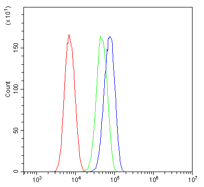
ARG42832 anti-MR1 antibody FACS image
Flow Cytometry: SiHa cells were blocked with 10% normal goat serum and then stained with ARG42832 anti-MR1 antibody (blue) at 1 µg/10^6 cells for 30 min at 20°C, followed by incubation with DyLight®488 labelled secondary antibody. Isotype control antibody (green) was Rabbit IgG (1 µg/10^6 cells) used under the same conditions. Unlabelled sample (red) was also used as a control.