ARG41772
anti-MOG / Myelin oligodendrocyte glycoprotein antibody
anti-MOG / Myelin oligodendrocyte glycoprotein antibody for Flow cytometry,IHC-Formalin-fixed paraffin-embedded sections,Western blot and Human,Mouse,Rat
Overview
| Product Description | Rabbit Polyclonal antibody recognizes MOG / Myelin oligodendrocyte glycoprotein |
|---|---|
| Tested Reactivity | Hu, Ms, Rat |
| Tested Application | FACS, IHC-P, WB |
| Host | Rabbit |
| Clonality | Polyclonal |
| Isotype | IgG |
| Target Name | MOG / Myelin oligodendrocyte glycoprotein |
| Antigen Species | Human |
| Immunogen | Synthetic peptide corresponding to a sequence of Human MOG / Myelin oligodendrocyte glycoprotein. (RVVHLYRNGKDQDGDQAPEYRGRTELLKDAIGEGK) |
| Conjugation | Un-conjugated |
| Alternate Names | BTNL11; BTN6; NRCLP7; MOGIG2; Myelin-oligodendrocyte glycoprotein |
Application Instructions
| Application Suggestion |
|
||||||||
|---|---|---|---|---|---|---|---|---|---|
| Application Note | IHC-P: Antigen Retrieval: Heat mediation was performed in Citrate buffer (pH 6.0, epitope retrieval solution) for 20 min. * The dilutions indicate recommended starting dilutions and the optimal dilutions or concentrations should be determined by the scientist. |
||||||||
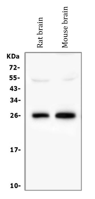
| Observed Size | ~ 26 kDa |
Properties
| Form | Liquid |
|---|---|
| Purification | Affinity purification with immunogen. |
| Buffer | 0.2% Na2HPO4, 0.9% NaCl, 0.05% Sodium azide and 4% Trehalose. |
| Preservative | 0.05% Sodium azide |
| Stabilizer | 4% Trehalose |
| Concentration | 0.5 mg/ml |
| Storage Instruction | For continuous use, store undiluted antibody at 2-8°C for up to a week. For long-term storage, aliquot and store at -20°C or below. Storage in frost free freezers is not recommended. Avoid repeated freeze/thaw cycles. Suggest spin the vial prior to opening. The antibody solution should be gently mixed before use. |
| Note | For laboratory research only, not for drug, diagnostic or other use. |
Bioinformation
| Database Links | |
|---|---|
| Gene Symbol | MOG |
| Gene Full Name | myelin oligodendrocyte glycoprotein |
| Background | The product of this gene is a membrane protein expressed on the oligodendrocyte cell surface and the outermost surface of myelin sheaths. Due to this localization, it is a primary target antigen involved in immune-mediated demyelination. This protein may be involved in completion and maintenance of the myelin sheath and in cell-cell communication. Alternatively spliced transcript variants encoding different isoforms have been identified. [provided by RefSeq, Jul 2008] |
| Function | Mediates homophilic cell-cell adhesion (By similarity). Minor component of the myelin sheath. May be involved in completion and/or maintenance of the myelin sheath and in cell-cell communication. [UniProt] |
| Cellular Localization | Isoform 1 and 5: Cell membrane; Multi-pass membrane protein. Isoform 2, 3, 4, 6, 7, 8 and 9: Cell membrane; Single-pass type I membrane protein. [UniProt] |
| Calculated MW | 28 kDa |
Images (6) Click the Picture to Zoom In
-
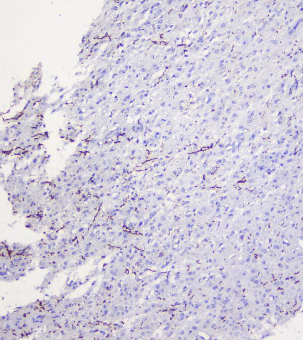
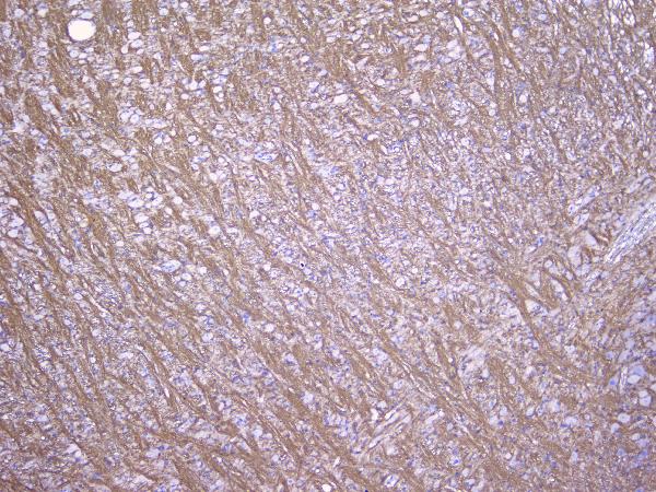
ARG41772 anti-MOG / Myelin oligodendrocyte glycoprotein antibody IHC-P image
Immunohistochemistry: Paraffin-embedded Human glioma tissue. Antigen Retrieval: Heat mediation was performed in Citrate buffer (pH 6.0, epitope retrieval solution) for 20 min. The tissue section was blocked with 10% goat serum. The tissue section was then stained with ARG41772 anti-MOG / Myelin oligodendrocyte glycoprotein antibody at 1 µg/ml dilution, overnight at 4°C.
-

ARG41772 anti-MOG / Myelin oligodendrocyte glycoprotein antibody WB image
Western blot: 50 µg of samples under reducing conditions. Rat brain and Mouse brain lysates stained with ARG41772 anti-MOG / Myelin oligodendrocyte glycoprotein antibody at 0.5 µg/ml dilution, overnight at 4°C.
-
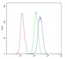
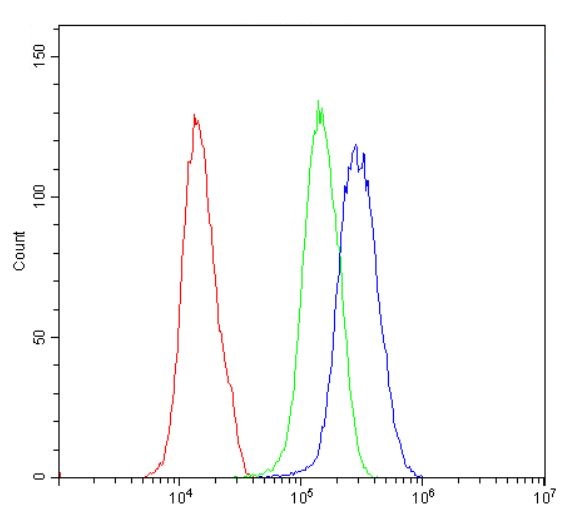
ARG41772 anti-MOG / Myelin oligodendrocyte glycoprotein antibody FACS image
Flow Cytometry: U251 cells were blocked with 10% normal goat serum and then stained with ARG41772 anti-MOG / Myelin oligodendrocyte glycoprotein antibody (blue) at 1 µg/10^6 cells for 30 min at 20°C, followed by incubation with DyLight®488 labelled secondary antibody. Isotype control antibody (green) was Rabbit IgG (1 µg/10^6 cells) used under the same conditions. Unlabelled sample (red) was also used as a control.
-
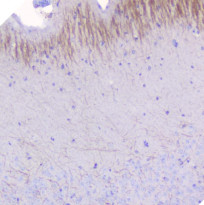
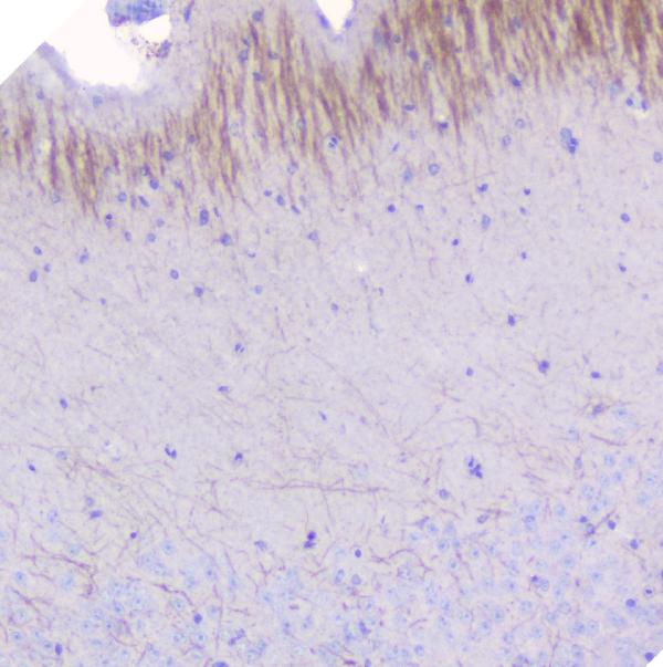
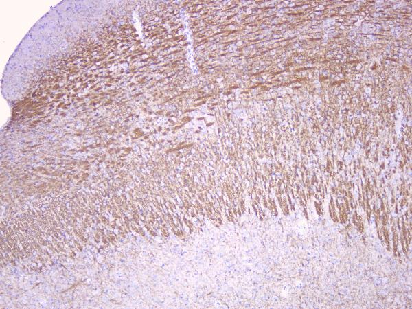
ARG41772 anti-MOG / Myelin oligodendrocyte glycoprotein antibody IHC-P image
Immunohistochemistry: Paraffin-embedded Mouse brain tissue. Antigen Retrieval: Heat mediation was performed in Citrate buffer (pH 6.0, epitope retrieval solution) for 20 min. The tissue section was blocked with 10% goat serum. The tissue section was then stained with ARG41772 anti-MOG / Myelin oligodendrocyte glycoprotein antibody at 1 µg/ml dilution, overnight at 4°C.
-
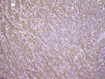
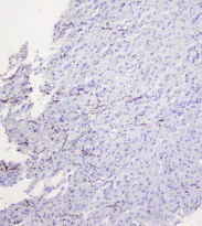
ARG41772 anti-MOG / Myelin oligodendrocyte glycoprotein antibody IHC-P image
Immunohistochemistry: Paraffin-embedded Rat brain tissue. Antigen Retrieval: Heat mediation was performed in Citrate buffer (pH 6.0, epitope retrieval solution) for 20 min. The tissue section was blocked with 10% goat serum. The tissue section was then stained with ARG41772 anti-MOG / Myelin oligodendrocyte glycoprotein antibody at 1 µg/ml dilution, overnight at 4°C.
-
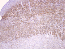
ARG41772 anti-MOG / Myelin oligodendrocyte glycoprotein antibody IHC-P image
Immunohistochemistry: Paraffin-embedded Rat brain tissue. Antigen Retrieval: Heat mediation was performed in Citrate buffer (pH 6.0, epitope retrieval solution) for 20 min. The tissue section was blocked with 10% goat serum. The tissue section was then stained with ARG41772 anti-MOG / Myelin oligodendrocyte glycoprotein antibody at 1 µg/ml dilution, overnight at 4°C.