ARG40476
anti-MMP16 antibody
anti-MMP16 antibody for Flow cytometry,ICC/IF,IHC-Formalin-fixed paraffin-embedded sections,Western blot and Human,Mouse
Overview
| Product Description | Rabbit Polyclonal antibody recognizes MMP16 |
|---|---|
| Tested Reactivity | Hu, Ms |
| Tested Application | FACS, ICC/IF, IHC-P, WB |
| Host | Rabbit |
| Clonality | Polyclonal |
| Isotype | IgG |
| Target Name | MMP16 |
| Antigen Species | Human |
| Immunogen | Recombinant protein corresponding to Y120-I296 of Human MMP16. |
| Conjugation | Un-conjugated |
| Alternate Names | MT-MMP 3; MT-MMP2; MT3-MMP; MTMMP3; MT-MMP3; C8orf57; EC 3.4.24.-; MMP-X2; Membrane-type matrix metalloproteinase 3; Membrane-type-3 matrix metalloproteinase; MMP-16; MT3MMP; Matrix metalloproteinase-16 |
Application Instructions
| Application Suggestion |
|
||||||||||
|---|---|---|---|---|---|---|---|---|---|---|---|
| Application Note | IHC-P: Antigen Retrieval: Heat mediation was performed in Citrate buffer (pH 6.0) for 20 min. * The dilutions indicate recommended starting dilutions and the optimal dilutions or concentrations should be determined by the scientist. |
Properties
| Form | Liquid |
|---|---|
| Purification | Affinity purification with immunogen. |
| Buffer | 0.2% Na2HPO4, 0.9% NaCl, 0.05% Sodium azide and 5% BSA. |
| Preservative | 0.05% Sodium azide |
| Stabilizer | 5% BSA |
| Concentration | 0.5 mg/ml |
| Storage Instruction | For continuous use, store undiluted antibody at 2-8°C for up to a week. For long-term storage, aliquot and store at -20°C or below. Storage in frost free freezers is not recommended. Avoid repeated freeze/thaw cycles. Suggest spin the vial prior to opening. The antibody solution should be gently mixed before use. |
| Note | For laboratory research only, not for drug, diagnostic or other use. |
Bioinformation
| Database Links | |
|---|---|
| Gene Symbol | MMP16 |
| Gene Full Name | matrix metallopeptidase 16 (membrane-inserted) |
| Background | Proteins of the matrix metalloproteinase (MMP) family are involved in the breakdown of extracellular matrix in normal physiological processes, such as embryonic development, reproduction, and tissue remodeling, as well as in disease processes, such as arthritis and metastasis. Most MMP's are secreted as inactive proproteins which are activated when cleaved by extracellular proteinases. The encoded protein activates MMP2 by cleavage. This gene was once referred to as MT-MMP2, but was renamed as MT-MMP3 or MMP16. [provided by RefSeq, Oct 2010] |
| Function | Endopeptidase that degrades various components of the extracellular matrix, such as collagen type III and fibronectin. Activates progelatinase A. Involved in the matrix remodeling of blood vessels. Isoform short cleaves fibronectin and also collagen type III, but at lower rate. It has no effect on type I, II, IV and V collagen. However, upon interaction with CSPG4, it may be involved in degradation and invasion of type I collagen by melanoma cells. [UniProt] |
| Cellular Localization | Isoform Long: Cell membrane; Single-pass type I membrane protein; Extracellular side. Note=Localized at the cell surface of melanoma cells. Isoform Short: Secreted, extracellular space, extracellular matrix. Cell surface. Note=Localized at the cell surface of melanoma cells. [UniProt] |
| Calculated MW | 70 kDa |
| PTM | The precursor is cleaved by a furin endopeptidase. [UniProt] |
Images (7) Click the Picture to Zoom In
-

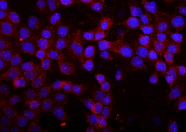
ARG40476 anti-MMP16 antibody ICC/IF image
Immunofluorescence: A431 cells were blocked with 10% goat serum and then stained with ARG40476 anti-MMP16 antibody (red) at 2 µg/ml dilution, overnight at 4°C. DAPI (blue) for nuclear staining.
-
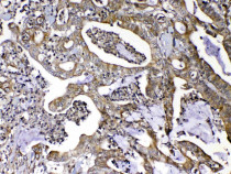
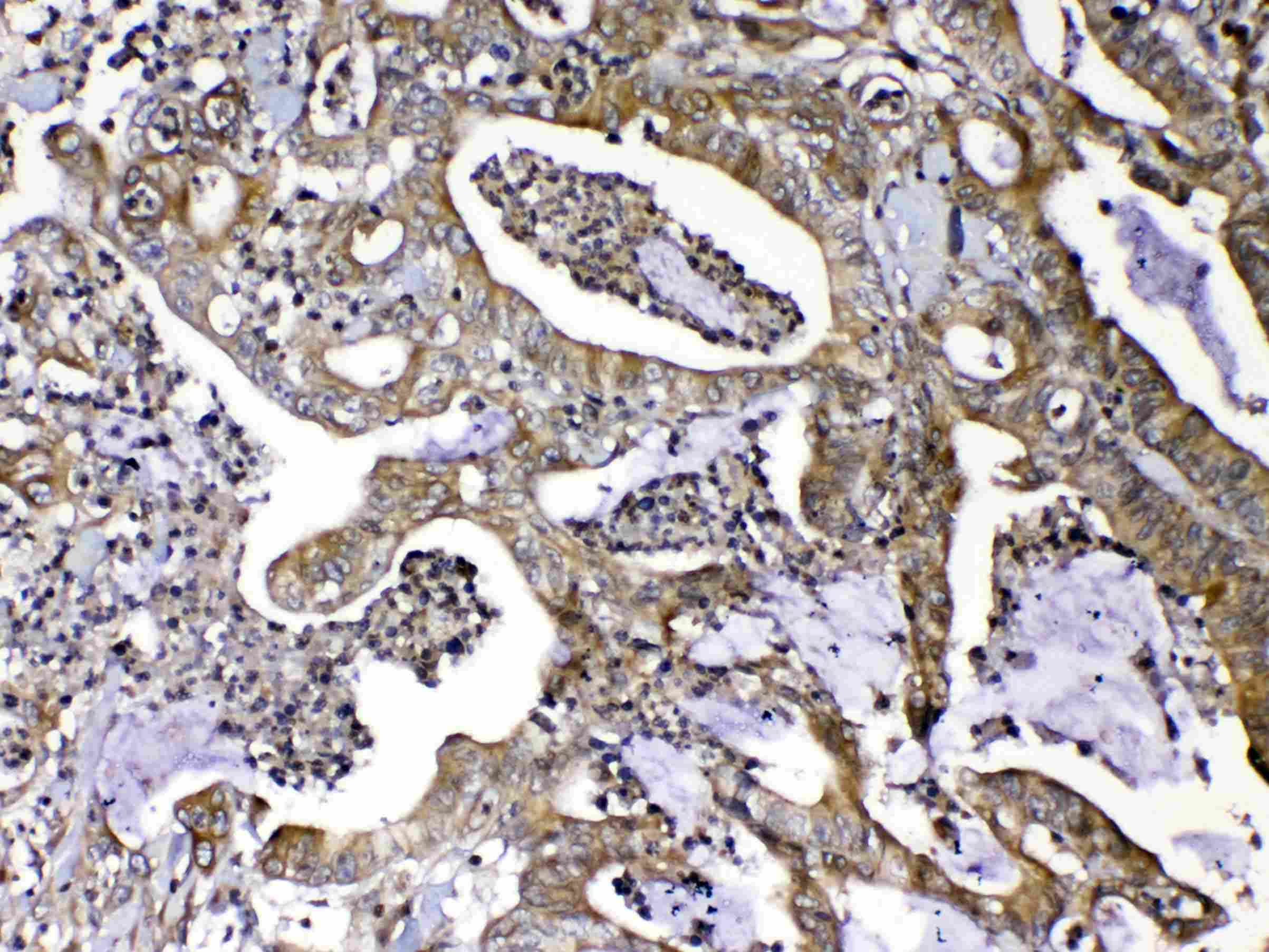
ARG40476 anti-MMP16 antibody IHC-P image
Immunohistochemistry: Paraffin-embedded Human colon cancer tissue. Antigen Retrieval: Heat mediation was performed in Citrate buffer (pH 6.0, epitope retrieval solution) for 20 min. The tissue section was blocked with 10% goat serum. The tissue section was then stained with ARG40476 anti-MMP16 antibody at 1 µg/ml, overnight at 4°C.
-
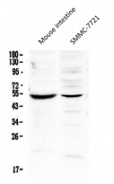
ARG40476 anti-MMP16 antibody WB image
Western blot: 50 µg of samples under reducing conditions. Mouse intestine and SMMC-7721 whole cell lysates stained with ARG40476 anti-MMP16 antibody at 0.5 µg/ml, overnight at 4°C.
-
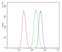
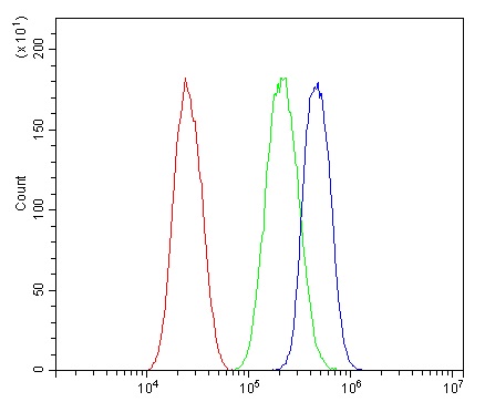
ARG40476 anti-MMP16 antibody FACS image
Flow Cytometry: Caco-2 cells were blocked with 10% normal goat serum and then stained with ARG40476 anti-MMP16 antibody (blue) at 1 µg/10^6 cells for 30 min at 20°C, followed by incubation with DyLight®488 labelled secondary antibody. Isotype control antibody (green) was rabbit IgG (1 µg/10^6 cells) used under the same conditions. Unlabelled sample (red) was also used as a control.
-

ARG40476 anti-MMP16 antibody IHC-P image
Immunohistochemistry: Paraffin-embedded Human lung cancer tissue. Antigen Retrieval: Heat mediation was performed in Citrate buffer (pH 6.0, epitope retrieval solution) for 20 min. The tissue section was blocked with 10% goat serum. The tissue section was then stained with ARG40476 anti-MMP16 antibody at 1 µg/ml, overnight at 4°C.
-
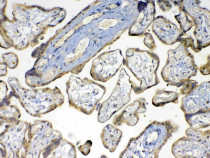
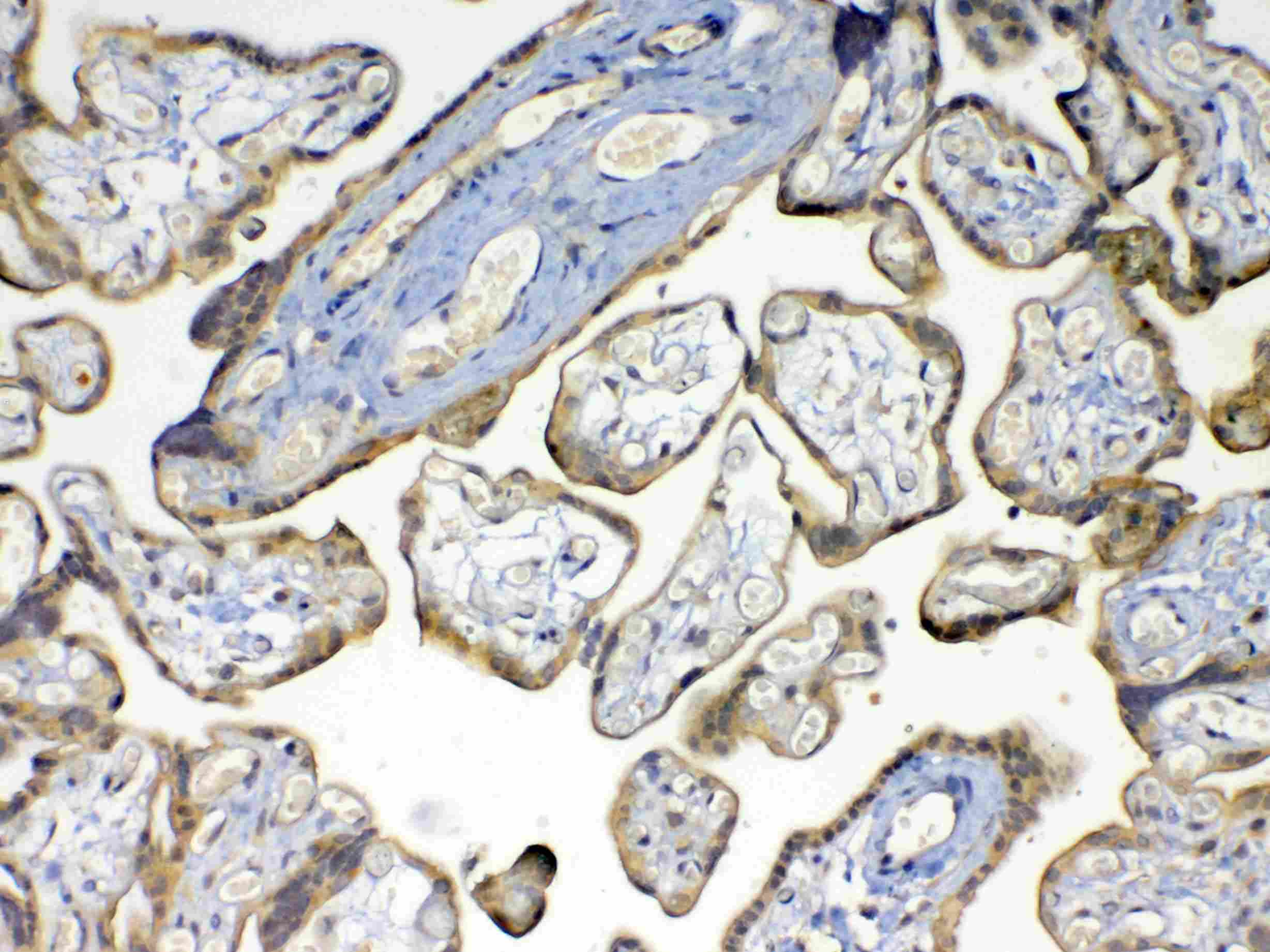
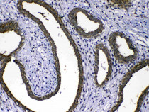
ARG40476 anti-MMP16 antibody IHC-P image
Immunohistochemistry: Paraffin-embedded Human placenta tissue. Antigen Retrieval: Heat mediation was performed in Citrate buffer (pH 6.0, epitope retrieval solution) for 20 min. The tissue section was blocked with 10% goat serum. The tissue section was then stained with ARG40476 anti-MMP16 antibody at 1 µg/ml, overnight at 4°C.
-
ARG40476 anti-MMP16 antibody IHC-P image
Immunohistochemistry: Paraffin-embedded Human mammary cancer tissue. Antigen Retrieval: Heat mediation was performed in Citrate buffer (pH 6.0, epitope retrieval solution) for 20 min. The tissue section was blocked with 10% goat serum. The tissue section was then stained with ARG40476 anti-MMP16 antibody at 1 µg/ml, overnight at 4°C.