ARG55847
anti-MICA antibody
anti-MICA antibody for Flow cytometry,ICC/IF,IHC-Formalin-fixed paraffin-embedded sections,Western blot and Human
Overview
| Product Description | Rabbit Polyclonal antibody recognizes MICA |
|---|---|
| Tested Reactivity | Hu |
| Tested Application | FACS, ICC/IF, IHC-P, WB |
| Host | Rabbit |
| Clonality | Polyclonal |
| Isotype | IgG |
| Target Name | MICA |
| Antigen Species | Human |
| Immunogen | KLH-conjugated synthetic peptide corresponding to aa. 68-97 (Center) of Human MICA. |
| Conjugation | Un-conjugated |
| Alternate Names | MHC class I polypeptide-related sequence A; PERB11.1; MIC-A |
Application Instructions
| Application Suggestion |
|
||||||||||
|---|---|---|---|---|---|---|---|---|---|---|---|
| Application Note | * The dilutions indicate recommended starting dilutions and the optimal dilutions or concentrations should be determined by the scientist. | ||||||||||
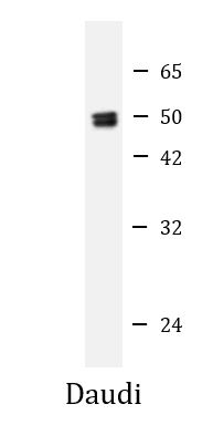
| Positive Control | Daudi | ||||||||||
| Observed Size | ~ 50 kDa |
Properties
| Form | Liquid |
|---|---|
| Purification | Purification with Protein A and immunogen peptide. |
| Buffer | PBS and 0.09% (W/V) Sodium azide |
| Preservative | 0.09% (W/V) Sodium azide |
| Storage Instruction | For continuous use, store undiluted antibody at 2-8°C for up to a week. For long-term storage, aliquot and store at -20°C or below. Storage in frost free freezers is not recommended. Avoid repeated freeze/thaw cycles. Suggest spin the vial prior to opening. The antibody solution should be gently mixed before use. |
| Note | For laboratory research only, not for drug, diagnostic or other use. |
Bioinformation
| Database Links |
Swiss-port # Q29983 Human MHC class I polypeptide-related sequence A |
|---|---|
| Gene Symbol | MICA |
| Gene Full Name | MHC class I polypeptide-related sequence A |
| Background | This gene encodes the highly polymorphic major histocompatability complex class I chain-related protein A. The protein product is expressed on the cell surface, although unlike canonical class I molecules it does not seem to associate with beta-2-microglobulin. It is a ligand for the NKG2-D type II integral membrane protein receptor. The protein functions as a stress-induced antigen that is broadly recognized by intestinal epithelial gamma delta T cells. Variations in this gene have been associated with susceptibility to psoriasis 1 and psoriatic arthritis, and the shedding of MICA-related antibodies and ligands is involved in the progression from monoclonal gammopathy of undetermined significance to multiple myeloma. Alternative splicing of this gene results in multiple transcript variants. [provided by RefSeq, Jan 2014] |
| Function | Seems to have no role in antigen presentation. Acts as a stress-induced self-antigen that is recognized by gamma delta T-cells. Ligand for the KLRK1/NKG2D receptor. Binding to KLRK1 leads to cell lysis. [UniProt] |
| Cellular Localization | Cell membrane; Single- pass type I membrane protein Cytoplasm Note=Expressed on the cell surface in gastric epithelium, endothelial cells and fibroblasts and in the cytoplasm in keratinocytes and monocytes. Infection with human adenovirus 5 suppresses cell surface expression due to the adenoviral E3-19K protein which causes retention in the endoplasmic reticulum |
| Calculated MW | 43 kDa |
| PTM | N-glycosylated. Glycosylation is not essential for interaction with KLRK1/NKG2D but enhances complex formation. Proteolytically cleaved and released from the cell surface of tumor cells which impairs KLRK1/NKG2D expression and T-cell activation. |
Images (4) Click the Picture to Zoom In
-
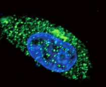
ARG55847 anti-MICA antibody ICC/IF image
Immunofluorescence: MDA-MB-231 cells stained with ARG55847 anti-MICA antibody (green). DAPI (blue) for nuclear staining.
-
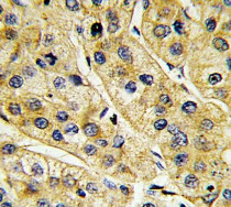
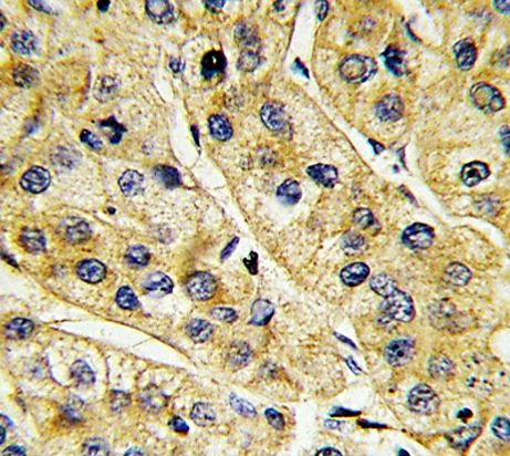
ARG55847 anti-MICA antibody IHC-P image
Immunohistochemistry: Formalin-fixed and paraffin-embedded Human hepatocarcinoma stained with ARG55847 anti-MICA antibody.
-
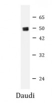
ARG55847 anti-MICA antibody WB image
Western blot: 20 µg of Daudi whole cell lysate stained with ARG55847 anti-MICA antibody at 1:1000 dilution.
-
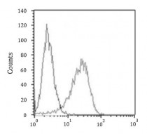
ARG55847 anti-MICA antibody FACS image
Flow Cytometry: SK-BR-3 cells stained with ARG55847 anti-MICA antibody (right histogram) at 1:25 dilution or isotype control antibody (left histogram), followed by incubation with Alexa Fluor® 488 labelled secondary antibody.