ARG41163
anti-MGEA5 / OGA antibody
anti-MGEA5 / OGA antibody for Flow cytometry,IHC-Formalin-fixed paraffin-embedded sections,Western blot and Human
Overview
| Product Description | Rabbit Polyclonal antibody recognizes MGEA5 / OGA |
|---|---|
| Tested Reactivity | Hu |
| Tested Application | FACS, IHC-P, WB |
| Host | Rabbit |
| Clonality | Polyclonal |
| Isotype | IgG |
| Target Name | MGEA5 / OGA |
| Antigen Species | Human |
| Immunogen | KLH-conjugated synthetic peptide between aa. 236-269 of Human MGEA5. |
| Conjugation | Un-conjugated |
| Alternate Names | Nuclear cytoplasmic O-GlcNAcase and acetyltransferase; N-acetyl-beta-D-glucosaminidase; MEA5; NCOAT; EC 3.2.1.-; Meningioma-expressed antigen 5; Beta-N-acetylhexosaminidase; Beta-hexosaminidase; EC 3.2.1.169; Protein O-GlcNAcase; Beta-N-acetylglucosaminidase; OGA; N-acetyl-beta-glucosaminidase |
Application Instructions
| Application Suggestion |
|
||||||||
|---|---|---|---|---|---|---|---|---|---|
| Application Note | IHC-P: Antigen Retrieval: Heat mediation was performed in Citrate buffer (pH 6.0). * The dilutions indicate recommended starting dilutions and the optimal dilutions or concentrations should be determined by the scientist. |
||||||||
| Positive Control | U-87 MG | ||||||||
| Observed Size | ~120-130 kda |
Properties
| Form | Liquid |
|---|---|
| Purification | Purification with Protein A and immunogen peptide. |
| Buffer | PBS and 0.09% (W/V) Sodium azide. |
| Preservative | 0.09% (W/V) Sodium azide |
| Storage Instruction | For continuous use, store undiluted antibody at 2-8°C for up to a week. For long-term storage, aliquot and store at -20°C or below. Storage in frost free freezers is not recommended. Avoid repeated freeze/thaw cycles. Suggest spin the vial prior to opening. The antibody solution should be gently mixed before use. |
| Note | For laboratory research only, not for drug, diagnostic or other use. |
Bioinformation
| Database Links | |
|---|---|
| Gene Symbol | MGEA5 |
| Gene Full Name | meningioma expressed antigen 5 (hyaluronidase) |
| Background | The dynamic modification of cytoplasmic and nuclear proteins by O-linked N-acetylglucosamine (O-GlcNAc) addition and removal on serine and threonine residues is catalyzed by OGT (MIM 300255), which adds O-GlcNAc, and MGEA5, a glycosidase that removes O-GlcNAc modifications (Gao et al., 2001 [PubMed 11148210]).[supplied by OMIM, Mar 2008] |
| Function | Isoform 1: Cleaves GlcNAc but not GalNAc from O-glycosylated proteins. Can use p-nitrophenyl-beta-GlcNAc and 4-methylumbelliferone-GlcNAc as substrates but not p-nitrophenyl-beta-GalNAc or p-nitrophenyl-alpha-GlcNAc (in vitro). Does not bind acetyl-CoA and does not have histone acetyltransferase activity. Isoform 3: Cleaves GlcNAc but not GalNAc from O-glycosylated proteins. Can use p-nitrophenyl-beta-GlcNAc as substrate but not p-nitrophenyl-beta-GalNAc or p-nitrophenyl-alpha-GlcNAc (in vitro), but has about six times lower specific activity than isoform 1. [UniProt] |
| Cellular Localization | Isoform 3: Nucleus. Isoform 1: Cytoplasm. [UniProt] |
| Calculated MW | 103 kDa |
| PTM | Proteolytically cleaved by caspase-3 during apoptosis. The fragments interact with each other; cleavage does not decrease enzyme activity. [UniProt] |
Images (3) Click the Picture to Zoom In
-
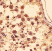
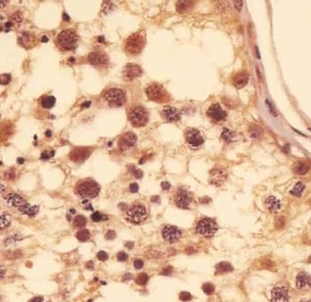
ARG41163 anti-MGEA5 / OGA antibody IHC-P image
Immunohistochemistry: Paraformaldehyde-fixed and paraffin-embedded Human testis tissue. Tissue was blocked with 3% BSA for 0.5 hour at room temperature. Antigen Retrieval: Heat mediation was performed in Citrate buffer (pH 6.0). Samples were stained with ARG41163 anti-MGEA5 / OGA antibody at 1:25 dilution for 1 hour at 37°C.
-
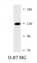
ARG41163 anti-MGEA5 / OGA antibody WB image
Western blot: 20 µg of U-87 MG cell lysate stained with ARG41163 anti-MGEA5 / OGA antibody at 1:2000 dilution.
-
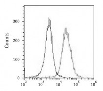
ARG41163 anti-MGEA5 / OGA antibody FACS image
Flow Cytometry: HeLa cells were fixed with 2% paraformaldehyde (10 min) and then permeabilized with 90% methanol for 10 min. The cells were then incubated in 2% BSA to block non-specific protein-protein interactions followed by ARG41163 anti-MGEA5 / OGA antibody (right histogram) at 1:25 dilution for 60 min at 37°C, followed by incubation with DyLight®488 labelled secondary antibody. Isotype control antibody (left histogram) was Rabbit IgG1 (1 µg/10^6 cells) used under the same conditions. Acquisition of > 10000 events was performed.