ARG44140
anti-MFAP3 antibody
anti-MFAP3 antibody for Flow cytometry,IHC-Formalin-fixed paraffin-embedded sections,Western blot and Human,Mouse,Rat
Overview
| Product Description | Rabbit Polyclonal recognizes MFAP3 |
|---|---|
| Tested Reactivity | Hu, Ms, Rat |
| Tested Application | FACS, IHC-P, WB |
| Host | Rabbit |
| Clonality | Polyclonal |
| Isotype | IgG |
| Target Name | MFAP3 |
| Antigen Species | Human |
| Immunogen | Human MFAP3 recombinant protein (Position: A19-N349). |
| Conjugation | Un-conjugated |
| Alternate Names | MFAP3; Microfibril Associated Protein 3; Microfibril-Associated Glycoprotein 3; Microfibrillar Associated Protein 3 |
Application Instructions
| Application Suggestion |
|
||||||||
|---|---|---|---|---|---|---|---|---|---|
| Application Note | The dilutions indicate recommended starting dilutions and the optimal dilutions or concentrations should be determined by the scientist. |
Properties
| Form | Liquid |
|---|---|
| Purification | Affinity purification with immunogen. |
| Buffer | 0.9% NaCl, 0.2% Na2HPO4, 0.05% Sodium azide and 4% Trehalose. |
| Preservative | 0.05% Sodium azide |
| Stabilizer | 4% Trehalose |
| Concentration | 0.5 mg/ml |
| Storage Instruction | For continuous use, store undiluted antibody at 2-8°C for up to a week. For long-term storage, aliquot and store at -20°C or below. Storage in frost free freezers is not recommended. Avoid repeated freeze/thaw cycles. Suggest spin the vial prior to opening. The antibody solution should be gently mixed before use. |
| Note | For laboratory research only, not for drug, diagnostic or other use. |
Bioinformation
| Database Links | |
|---|---|
| Gene Symbol | MFAP3 |
| Gene Full Name | Microfibril Associated Protein 3 |
| Background | Predicted to be located in extracellular region. Predicted to be active in cytoplasm; nucleus; and plasma membrane. |
| Function | Component of the elastin-associated microfibrils. |
| Cellular Localization | Cell membrane, Membrane |
| Calculated MW | 40 kDa |
| PTM | Disulfide bond, Glycoprotein |
Images (5) Click the Picture to Zoom In
-
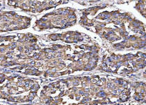
ARG44140 anti-MFAP3 antibody IHC-P image
Immunohistochemistry: Human liver cancer stained with ARG44140 anti-MFAP3 antibody at 2 μg/ml dilution.
-
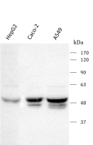
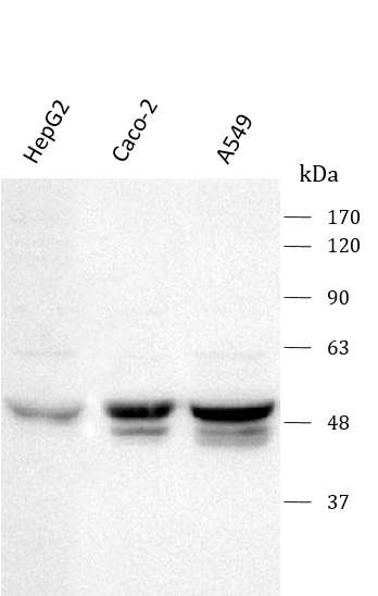
ARG44140 anti-MFAP3 antibody WB image
Western blot: HepG2, Caco-2 and A549 stained with ARG44140 anti-MFAP3 antibody at 0.5 μg/ml dilution.
-
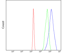
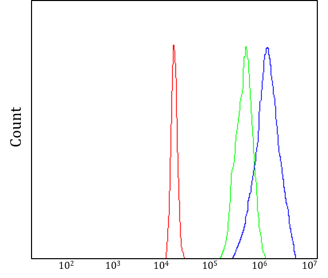
ARG44140 anti-MFAP3 antibody FACS image
Flow Cytometry: 293T stained with ARG44140 anti-MFAP3 antibody at 1 µg/10^6 cells dilution.
-
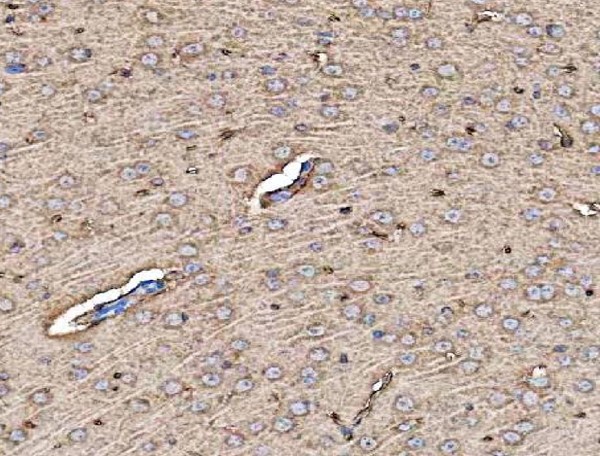
ARG44140 anti-MFAP3 antibody IHC-P image
Immunohistochemistry: Rat brain stained with ARG44140 anti-MFAP3 antibody at 2 μg/ml dilution.
-
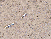
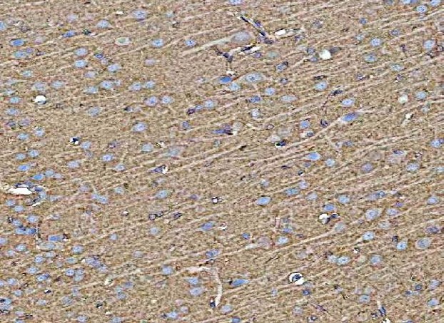
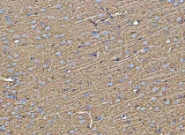
ARG44140 anti-MFAP3 antibody IHC-P image
Immunohistochemistry: Mouse brain stained with ARG44140 anti-MFAP3 antibody at 2 μg/ml dilution.