ARG44191
anti-MFAP1 antibody
anti-MFAP1 antibody for Flow cytometry,ICC/IF,Western blot and Human,Mouse
Overview
| Product Description | Rabbit Polyclonal antibody recognizes MFAP1 |
|---|---|
| Tested Reactivity | Hu, Ms |
| Tested Application | FACS, ICC/IF, WB |
| Host | Rabbit |
| Clonality | Polyclonal |
| Isotype | IgG |
| Target Name | MFAP1 |
| Antigen Species | Human |
| Immunogen | Recombinant protein of Human MFAP1 |
| Conjugation | Un-conjugated |
| Protein Full Name | Microfibrillar-associated protein 1 |
| Alternate Names | MFAP1; Microfibril Associated Protein 1; AMP; Microfibrillar Associated Protein 1; Microfibrillar-Associated Protein 1; Spliceosome B Complex Protein MFAP1 |
Application Instructions
| Application Suggestion |
|
||||||||
|---|---|---|---|---|---|---|---|---|---|
| Application Note | * The dilutions indicate recommended starting dilutions and the optimal dilutions or concentrations should be determined by the scientist. |
Properties
| Form | Liquid |
|---|---|
| Purification | Affinity purification with immunogen. |
| Buffer | 0.9% NaCl, 0.2% Na2HPO4, 0.05% Sodium azide and 5% BSA. |
| Preservative | 0.05% Sodium azide |
| Stabilizer | 5% BSA |
| Concentration | 0.5 mg/ml |
| Storage Instruction | For continuous use, store undiluted antibody at 2-8°C for up to a week. For long-term storage, aliquot and store at -20°C or below. Storage in frost free freezers is not recommended. Avoid repeated freeze/thaw cycles. Suggest spin the vial prior to opening. The antibody solution should be gently mixed before use. |
| Note | For laboratory research only, not for drug, diagnostic or other use. |
Bioinformation
| Database Links |
Swiss-port # P55081 Human Microfibrillar-associated protein 1 |
|---|---|
| Gene Symbol | MFAP1 |
| Gene Full Name | Microfibril Associated Protein 1 |
| Background | Enables RNA binding activity. Involved in mRNA splicing, via spliceosome. Located in centrosome; microfibril; and nucleoplasm. Part of U2-type precatalytic spliceosome. |
| Function | Involved in pre-mRNA splicing as a component of the spliceosome. |
| Cellular Localization | Nucleus, Spliceosome |
| Calculated MW | 52 kDa |
| PTM | Acetylation, Isopeptide bond, Phosphoprotein, Ubl conjugation |
Images (4) Click the Picture to Zoom In
-
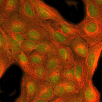
ARG44191 anti-MFAP1 antibody ICC/IF image
Immunofluorescence: U2OS stained with ARG44191 anti-MFAP1 antibody at 5 μg/mL dilution.
-
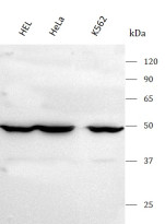
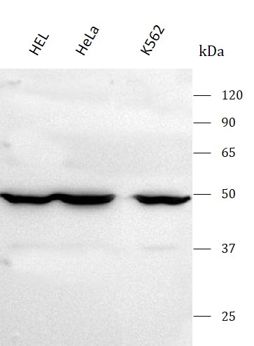
ARG44191 anti-MFAP1 antibody WB image
Western blot: HEL, Hela and K562 stained with ARG44191 anti-MFAP1 antibody at 0.5 μg/mL dilution.
-
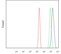
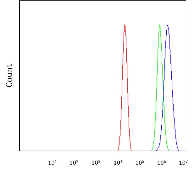
ARG44191 anti-MFAP1 antibody FACS image
Flow Cytometry: A431 stained with ARG44191 anti-MFAP1 antibody at 1 μg/1x10^6 cells dilution.
-
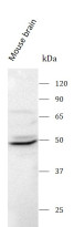
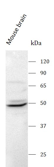
ARG44191 anti-MFAP1 antibody WB image
Western blot: Mouse brain stained with ARG44191 anti-MFAP1 antibody at 0.5 μg/mL dilution.