ARG22275
anti-MDC1 antibody [P2B11]
anti-MDC1 antibody [P2B11] for ICC/IF,Western blot and Human,Mouse,Bovine,Chimpanzee
Overview
| Product Description | Mouse Monoclonal antibody [P2B11] recognizes MDC1 |
|---|---|
| Tested Reactivity | Hu, Ms, Bov, Chimp |
| Tested Application | ICC/IF, WB |
| Specificity | Detects ~184kDa. This antibody recognizes MDC1 at and around the N-terminus. |
| Host | Mouse |
| Clonality | Monoclonal |
| Clone | P2B11 |
| Isotype | IgG1 |
| Target Name | MDC1 |
| Antigen Species | Mouse |
| Immunogen | GST-tagged recombinant protein around the N-terminus of Mouse MDC1 |
| Conjugation | Un-conjugated |
| Alternate Names | Nuclear factor with BRCT domains 1; Mediator of DNA damage checkpoint protein 1; NFBD1 |
Application Instructions
| Application Suggestion |
|
||||||
|---|---|---|---|---|---|---|---|
| Application Note | * The dilutions indicate recommended starting dilutions and the optimal dilutions or concentrations should be determined by the scientist. |
Properties
| Form | Liquid |
|---|---|
| Purification | Purification with Protein G. |
| Buffer | PBS (pH 7.4), 0.09% Sodium azide and 50% Glycerol |
| Preservative | 0.09% Sodium azide |
| Stabilizer | 50% Glycerol |
| Concentration | 1 mg/ml |
| Storage Instruction | For continuous use, store undiluted antibody at 2-8°C for up to a week. For long-term storage, aliquot and store at -20°C. Storage in frost free freezers is not recommended. Avoid repeated freeze/thaw cycles. Suggest spin the vial prior to opening. The antibody solution should be gently mixed before use. |
| Note | For laboratory research only, not for drug, diagnostic or other use. |
Bioinformation
| Database Links |
Swiss-port # Q14676 Human Mediator of DNA damage checkpoint protein 1 Swiss-port # Q5PSV9 Mouse Mediator of DNA damage checkpoint protein 1 |
|---|---|
| Gene Symbol | Mdc1 |
| Gene Full Name | mediator of DNA damage checkpoint 1 |
| Background | The protein encoded by this gene contains an N-terminal forkhead domain, two BRCA1 C-terminal (BRCT) motifs and a central domain with 13 repetitions of an approximately 41-amino acid sequence. The encoded protein is required to activate the intra-S phase and G2/M phase cell cycle checkpoints in response to DNA damage. This nuclear protein interacts with phosphorylated histone H2AX near sites of DNA double-strand breaks through its BRCT motifs, and facilitates recruitment of the ATM kinase and meiotic recombination 11 protein complex to DNA damage foci. [provided by RefSeq, Jul 2008] |
| Function | Required for checkpoint mediated cell cycle arrest in response to DNA damage within both the S phase and G2/M phases of the cell cycle. May serve as a scaffold for the recruitment of DNA repair and signal transduction proteins to discrete foci of DNA damage marked by 'Ser-139' phosphorylation of histone H2AFX. Also required for downstream events subsequent to the recruitment of these proteins. These include phosphorylation and activation of the ATM, CHEK1 and CHEK2 kinases, and stabilization of TP53 and apoptosis. ATM and CHEK2 may also be activated independently by a parallel pathway mediated by TP53BP1. [UniProt] |
| Cellular Localization | Nucleus |
| Calculated MW | 227 kDa |
| PTM | Phosphorylated upon exposure to ionizing radiation (IR), ultraviolet radiation (UV), and hydroxyurea (HU). Phosphorylation in response to IR requires ATM, NBN, and possibly CHEK2. Also phosphorylated during the G2/M phase of the cell cycle and during activation of the mitotic spindle checkpoint. Phosphorylation at Thr-4 by ATM stabilizes and enhances homodimerization via the FHA domain. Sumoylation at Lys-1840 by PIAS4 following DNA damage promotes ubiquitin-mediated degradation. Ubiquitinated by RNF4, leading to proteasomal degradation; undergoes 'Lys-48'-linked polyubiquitination. |
Images (3) Click the Picture to Zoom In
-

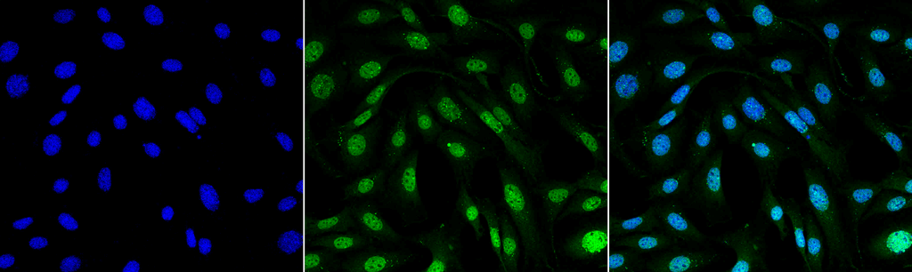
ARG22275 anti-MDC1 antibody [P2B11] ICC/IF image
Immunofluorescence: NIH 3T3 cells. Fixation: 4% Formaldehyde for 15 min at RT. Primary Antibody: ARG22275 anti-MDC1 antibody [P2B11] at 1:100 for 60 min at RT. Secondary Antibody: Goat anti-Mouse ATTO 488 at 1:100 for 60 min at RT. Counterstain: DAPI (blue) nuclear stain at 1:5000 for 5 min RT. Magnification: 60X. Left: DAPI (blue) nuclear stain. Middle: ARG22275 anti-MDC1 antibody [P2B11]. Right: Composite.
-
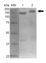
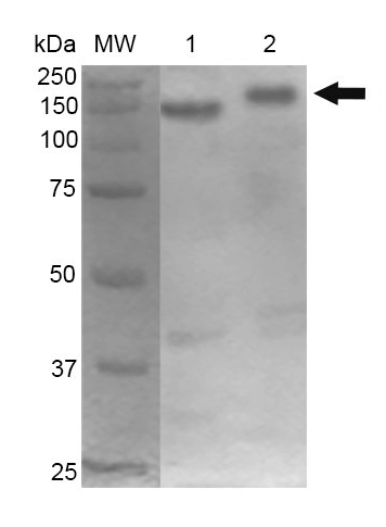
ARG22275 anti-MDC1 antibody [P2B11] WB image
Western blot: 10 µg of 1) Mouse Cortex, and 2) Cerebellum. Block: 5% Skim Milk in 1X TBST. Primary Antibody: ARG22275 anti-MDC1 antibody [P2B11] at 1:1000 for 2 hours RT. Secondary Antibody: Goat anti-Mouse HRP:IgG at 1:2000 for 60 min at RT. Color Development: ECL solution for 5 min in RT.
-

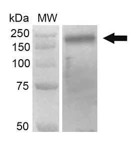
ARG22275 anti-MDC1 antibody [P2B11] WB image
Western blot: 30 µg of Human 293Trap cell lysates. Block: 5% Skim Milk in 1X TBST. Primary Antibody: ARG22275 anti-MDC1 antibody [P2B11] at 1:1000 for 2 hours RT. Secondary Antibody: Goat anti-Mouse HRP:IgG at 1:2000 for 60 min at RT. Color Development: ECL solution for 5 min in RT.