ARG59031
anti-MCM7 antibody
anti-MCM7 antibody for Flow cytometry,ICC/IF,IHC-Formalin-fixed paraffin-embedded sections,Western blot and Human,Mouse,Rat
Overview
| Product Description | Rabbit Polyclonal antibody recognizes MCM7 |
|---|---|
| Tested Reactivity | Hu, Ms, Rat |
| Tested Application | FACS, ICC/IF, IHC-P, WB |
| Host | Rabbit |
| Clonality | Polyclonal |
| Isotype | IgG |
| Target Name | MCM7 |
| Antigen Species | Human |
| Immunogen | Recombinant protein corresponding to D526-V719 of Human MCM7. |
| Conjugation | Un-conjugated |
| Alternate Names | PNAS146; CDC47 homolog; DNA replication licensing factor MCM7; MCM2; P1.1-MCM3; EC 3.6.4.12; CDC47; P1CDC47; P85MCM; PPP1R104 |
Application Instructions
| Application Suggestion |
|
||||||||||
|---|---|---|---|---|---|---|---|---|---|---|---|
| Application Note | IHC-P: Antigen Retrieval: Heat mediation was performed in Citrate buffer (pH 6.0) for 20 min. * The dilutions indicate recommended starting dilutions and the optimal dilutions or concentrations should be determined by the scientist. |
Properties
| Form | Liquid |
|---|---|
| Purification | Affinity purification with immunogen. |
| Buffer | 0.2% Na2HPO4, 0.9% NaCl, 0.05% Sodium azide and 5% BSA. |
| Preservative | 0.05% Sodium azide |
| Stabilizer | 5% BSA |
| Concentration | 0.5 mg/ml |
| Storage Instruction | For continuous use, store undiluted antibody at 2-8°C for up to a week. For long-term storage, aliquot and store at -20°C or below. Storage in frost free freezers is not recommended. Avoid repeated freeze/thaw cycles. Suggest spin the vial prior to opening. The antibody solution should be gently mixed before use. |
| Note | For laboratory research only, not for drug, diagnostic or other use. |
Bioinformation
| Database Links |
Swiss-port # P33993 Human DNA replication licensing factor MCM7 Swiss-port # Q61881 Mouse DNA replication licensing factor MCM7 |
|---|---|
| Gene Symbol | MCM7 |
| Gene Full Name | minichromosome maintenance complex component 7 |
| Background | The protein encoded by this gene is one of the highly conserved mini-chromosome maintenance proteins (MCM) that are essential for the initiation of eukaryotic genome replication. The hexameric protein complex formed by the MCM proteins is a key component of the pre-replication complex (pre_RC) and may be involved in the formation of replication forks and in the recruitment of other DNA replication related proteins. The MCM complex consisting of this protein and MCM2, 4 and 6 proteins possesses DNA helicase activity, and may act as a DNA unwinding enzyme. Cyclin D1-dependent kinase, CDK4, is found to associate with this protein, and may regulate the binding of this protein with the tumorsuppressor protein RB1/RB. Alternatively spliced transcript variants encoding distinct isoforms have been reported. [provided by RefSeq, Jul 2008] |
| Function | Acts as component of the MCM2-7 complex (MCM complex) which is the putative replicative helicase essential for 'once per cell cycle' DNA replication initiation and elongation in eukaryotic cells. The active ATPase sites in the MCM2-7 ring are formed through the interaction surfaces of two neighboring subunits such that a critical structure of a conserved arginine finger motif is provided in trans relative to the ATP-binding site of the Walker A box of the adjacent subunit. The six ATPase active sites, however, are likely to contribute differentially to the complex helicase activity. Required for S-phase checkpoint activation upon UV-induced damage. [UniProt] |
| Cellular Localization | Nucleus. [UniProt] |
| Calculated MW | 81 kDa |
| PTM | O-glycosylated (O-GlcNAcylated), in a cell cycle-dependent manner. [UniProt] |
Images (11) Click the Picture to Zoom In
-
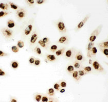
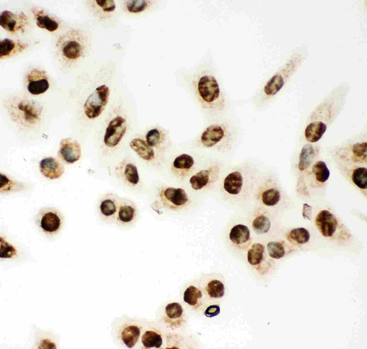
ARG59031 anti-MCM7 antibody ICC image
Immunocytochemistry: A549 cells stained with ARG59031 anti-MCM7 antibody.
-
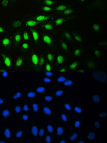
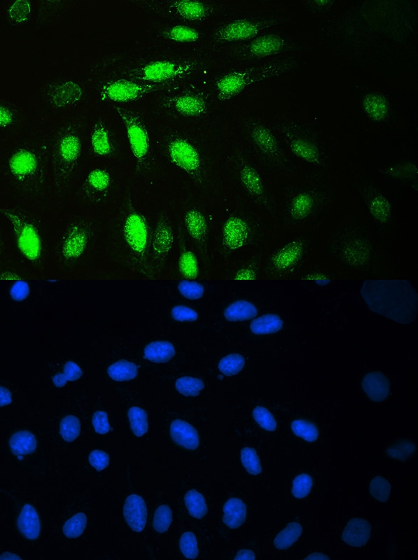
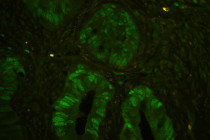
ARG59031 anti-MCM7 antibody ICC/IF image
Immunofluorescence: U2OS cells were blocked with 10% goat serum and then stained with ARG59031 anti-MCM7 antibody (green) at 2 µg/ml dilution, overnight at 4°C. DAPI (blue) for nuclear staining.
-
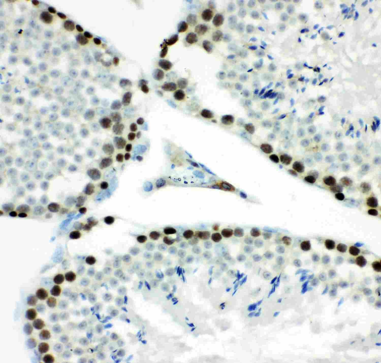
ARG59031 anti-MCM7 antibody IHC-P image
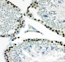
Immunohistochemistry: Paraffin-embedded Mouse testis tissue stained with ARG59031 anti-MCM7 antibody.
-
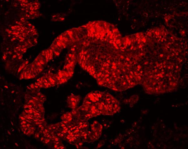
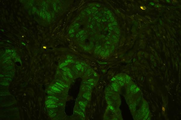
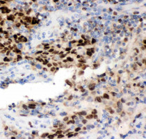
ARG59031 anti-MCM7 antibody IHC-P image
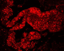
Immunohistochemistry: Paraffin-embedded Human colon cancer tissue. Antigen Retrieval: Heat mediation was performed in Citrate buffer (pH 6.0) for 20 min. The tissue section was blocked with 10% goat serum. The tissue section was then stained with ARG59031 anti-MCM7 antibody (red) at 2 µg/ml dilution, overnight at 4°C.
-
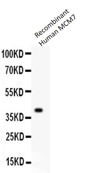
ARG59031 anti-MCM7 antibody WB image
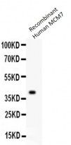
Western blot: 0.5 ng of Recombinant Human MCM7 Protein stained with ARG59031 anti-MCM7 antibody at 0.5 µg/ml.
-
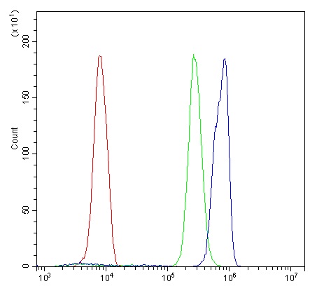
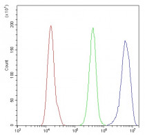
ARG59031 anti-MCM7 antibody FACS image
Flow Cytometry: U937 cells were blocked with 10% normal goat serum and then stained with ARG59031 anti-MCM7 antibody (blue) at 1 µg/10^6 cells for 30 min at 20°C, followed by incubation with DyLight®488 labelled secondary antibody. Isotype control antibody (green) was rabbit IgG (1 µg/10^6 cells) used under the same conditions. Unlabelled sample (red) was also used as a control.
-
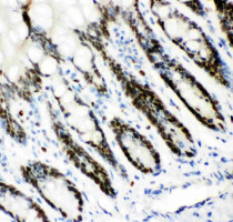
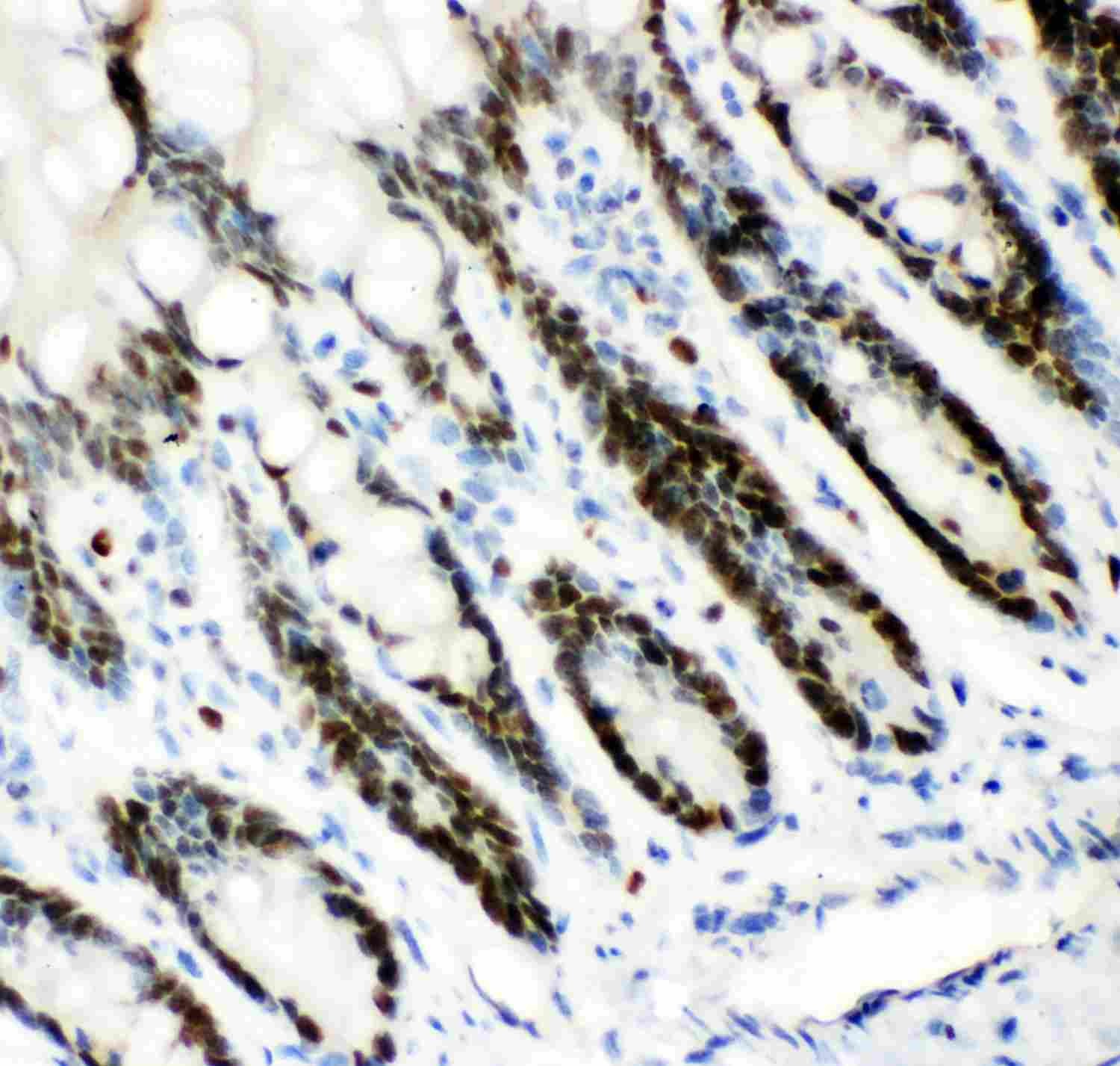
ARG59031 anti-MCM7 antibody IHC-P image
Immunohistochemistry: Paraffin-embedded Rat intestine tissue stained with ARG59031 anti-MCM7 antibody.
-
ARG59031 anti-MCM7 antibody IHC-P image
Immunohistochemistry: Paraffin-embedded Human colon cancer tissue. Antigen Retrieval: Heat mediation was performed in Citrate buffer (pH 6.0) for 20 min. The tissue section was blocked with 10% goat serum. The tissue section was then stained with ARG59031 anti-MCM7 antibody (green) at 2 µg/ml dilution, overnight at 4°C.
-
ARG59031 anti-MCM7 antibody IHC-P image
Immunohistochemistry: Paraffin-embedded Human lung cancer tissue stained with ARG59031 anti-MCM7 antibody.
-
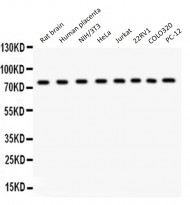
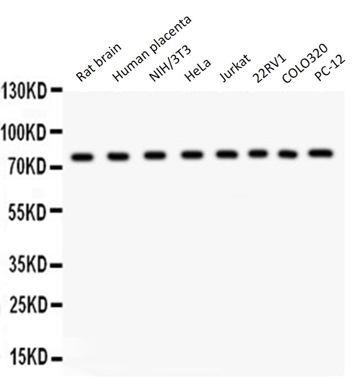
ARG59031 anti-MCM7 antibody WB image
Western blot: 50 µg of Rat brain, 50 µg of Human placenta, 40 µg of NIH/3T3, 40 µg of HeLa, 40 µg of Jurkat, 40 µg of 22RV1, 40 µg of COLO320 and 40 µg of PC-12 lysates stained with ARG59031 anti-MCM7 antibody at 0.5 µg/ml dilution.
-
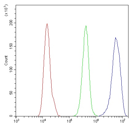
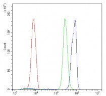
ARG59031 anti-MCM7 antibody FACS image
Flow Cytometry: A431 cells were blocked with 10% normal goat serum and then stained with ARG59031 anti-MCM7 antibody (blue) at 1 µg/10^6 cells for 30 min at 20°C, followed by incubation with DyLight®488 labelled secondary antibody. Isotype control antibody (green) was rabbit IgG (1 µg/10^6 cells) used under the same conditions. Unlabelled sample (red) was also used as a control.