ARG66305
anti-MAPKAPK2 / MK2 antibody
anti-MAPKAPK2 / MK2 antibody for IHC-Formalin-fixed paraffin-embedded sections,Immunoprecipitation,Western blot and Human,Mouse
Overview
| Product Description | Goat Polyclonal antibody recognizes MAPKAPK2 / MK2 |
|---|---|
| Tested Reactivity | Hu, Ms |
| Predict Reactivity | Cow, Rat, Dog |
| Tested Application | IHC-P, IP, WB |
| Host | Goat |
| Clonality | Polyclonal |
| Isotype | IgG |
| Target Name | MAPKAPK2 / MK2 |
| Antigen Species | Human |
| Immunogen | Synthetic peptide with sequence C-SRVLKEDKERWED, from the internal region (near C-terminus) of Human MAPKAPK2. (NP_004750.1; NP_116584.2) |
| Conjugation | Un-conjugated |
| Alternate Names | MAPKAP-K2; MAPK-activated protein kinase 2; MK-2; MAPKAP kinase 2; MAP kinase-activated protein kinase 2; MK2; EC 2.7.11.1; MAPKAPK-2 |
Application Instructions
| Application Suggestion |
|
||||||||
|---|---|---|---|---|---|---|---|---|---|
| Application Note | WB: Recommend incubate at RT for 1h. IHC-P: Antigen Retrieval: Steam tissue section in Citrate buffer (pH 6.0). * The dilutions indicate recommended starting dilutions and the optimal dilutions or concentrations should be determined by the scientist. |
Properties
| Form | Liquid |
|---|---|
| Purification | Affinity purified |
| Buffer | Tris saline (pH 7.3), 0.02% Sodium azide and 0.5% BSA. |
| Preservative | 0.02% Sodium azide |
| Stabilizer | 0.5% BSA |
| Concentration | 0.5 mg/ml |
| Storage Instruction | For continuous use, store undiluted antibody at 2-8°C for up to a week. For long-term storage, aliquot and store at -20°C or below. Storage in frost free freezers is not recommended. Avoid repeated freeze/thaw cycles. Suggest spin the vial prior to opening. The antibody solution should be gently mixed before use. |
| Note | For laboratory research only, not for drug, diagnostic or other use. |
Bioinformation
| Database Links |
Swiss-port # P49137 Human MAP kinase-activated protein kinase 2 Swiss-port # P49138 Mouse MAP kinase-activated protein kinase 2 |
|---|---|
| Gene Symbol | MAPKAPK2 |
| Gene Full Name | mitogen-activated protein kinase-activated protein kinase 2 |
| Background | This gene encodes a member of the Ser/Thr protein kinase family. This kinase is regulated through direct phosphorylation by p38 MAP kinase. In conjunction with p38 MAP kinase, this kinase is known to be involved in many cellular processes including stress and inflammatory responses, nuclear export, gene expression regulation and cell proliferation. Heat shock protein HSP27 was shown to be one of the substrates of this kinase in vivo. Two transcript variants encoding two different isoforms have been found for this gene. [provided by RefSeq, Jul 2008] |
| Function | Stress-activated serine/threonine-protein kinase involved in cytokines production, endocytosis, reorganization of the cytoskeleton, cell migration, cell cycle control, chromatin remodeling, DNA damage response and transcriptional regulation. Following stress, it is phosphorylated and activated by MAP kinase p38-alpha/MAPK14, leading to phosphorylation of substrates. Phosphorylates serine in the peptide sequence, Hyd-X-R-X(2)-S, where Hyd is a large hydrophobic residue. Phosphorylates ALOX5, CDC25B, CDC25C, ELAVL1, HNRNPA0, HSF1, HSP27/HSPB1, KRT18, KRT20, LIMK1, LSP1, PABPC1, PARN, PDE4A, RCSD1, RPS6KA3, TAB3 and TTP/ZFP36. Mediates phosphorylation of HSP27/HSPB1 in response to stress, leading to dissociate HSP27/HSPB1 from large small heat-shock protein (sHsps) oligomers and impair their chaperone activities and ability to protect against oxidative stress effectively. Involved in inflammatory response by regulating tumor necrosis factor (TNF) and IL6 production post-transcriptionally: acts by phosphorylating AU-rich elements (AREs)-binding proteins ELAVL1, HNRNPA0, PABPC1 and TTP/ZFP36, leading to regulate the stability and translation of TNF and IL6 mRNAs. Phosphorylation of TTP/ZFP36, a major post-transcriptional regulator of TNF, promotes its binding to 14-3-3 proteins and reduces its ARE mRNA affinity leading to inhibition of dependent degradation of ARE-containing transcript. Also involved in late G2/M checkpoint following DNA damage through a process of post-transcriptional mRNA stabilization: following DNA damage, relocalizes from nucleus to cytoplasm and phosphorylates HNRNPA0 and PARN, leading to stabilize GADD45A mRNA. Involved in toll-like receptor signaling pathway (TLR) in dendritic cells: required for acute TLR-induced macropinocytosis by phosphorylating and activating RPS6KA3. [UniProt] |
| Calculated MW | 46 kDa |
| PTM | Sumoylation inhibits the protein kinase activity. Phosphorylated and activated by MAP kinase p38-alpha/MAPK14 at Thr-222, Ser-272 and Thr-334. [UniProt] |
Images (4) Click the Picture to Zoom In
-
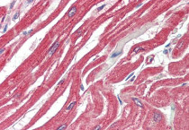
ARG66305 anti-MAPKAPK2 / MK2 antibody IHC-P image
Immunohistochemistry: Paraffin-embedded Human heart tissue. Antigen Retrieval: Steam tissue section in Citrate buffer (pH 6.0). The tissue section was stained with ARG66305 anti-MAPKAPK2 / MK2 antibody at 5 µg/ml dilution followed by AP-staining.
-
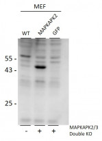
ARG66305 anti-MAPKAPK2 / MK2 antibody WB image
Western blot: 35 µg of MEF lysates stained with ARG66305 anti-MAPKAPK2 / MK2 antibody at 0.5 µg/ml dilution, for 2 hours at RT. Double KO mice in second and third lanes rescued by introduction of MAPKAPK2 gene in second lane.
-
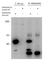
ARG66305 anti-MAPKAPK2 / MK2 antibody IP image
Immunoprecipitation: Lysates of MAPKAPK2 / MAPKAPK3 double knockout MEFs, with (lane 3) or without (lane 4) rescued MAPKAPK2 expression through retroviral transduction. The corresponding lysates (Lane 1 and 2) were analyzed in parallel in this Western blot labelled with Rabbit anti-MAPKAPK2 / MK2 antibody.
-
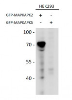
ARG66305 anti-MAPKAPK2 / MK2 antibody WB image
Western blot: HEK293 overexpressing Mouse MAPKAPK2 fused to GFP or overexpressing MAPKAPK5 fused to GFP. The blots were stained with ARG66305 anti-MAPKAPK2 / MK2 antibody at 0.5 µg/ml dilution and incubated at RT for 1 hour.


