ARG11033
anti-MAP2ab antibody [5H11]
anti-MAP2ab antibody [5H11] for ICC/IF,IHC-Frozen sections,Western blot and Human,Mouse,Rat,Bovine
Controls and Markers antibody; Neuroscience antibody; Signaling Transduction antibody; Neuron Marker antibody; Mature Neuron Marker antibody; Neurite Marker antibody
Overview
| Product Description | Mouse Monoclonal antibody [5H11] recognizes MAP2ab |
|---|---|
| Tested Reactivity | Hu, Ms, Rat, Bov |
| Tested Application | ICC/IF, IHC-Fr, WB |
| Specificity | This antibody was raised against purified full length bovine brain MAP2 and the epitope was mapped to aa. 1057-1507 of the human MAP2 sequence, so it reacts to MAP2a and MAP2b proteins. |
| Host | Mouse |
| Clonality | Monoclonal |
| Clone | 5H11 |
| Isotype | IgG2b |
| Target Name | MAP2ab |
| Antigen Species | Human |
| Immunogen | Full length purified Bovine MAP2. |
| Epitope | aa. 1057-1507 of the human MAP2 sequence. |
| Conjugation | Un-conjugated |
| Alternate Names | MAP2A; Microtubule-associated protein 2; MAP2B; MAP-2 |
Application Instructions
| Application Suggestion |
|
||||||||
|---|---|---|---|---|---|---|---|---|---|
| Application Note | * The dilutions indicate recommended starting dilutions and the optimal dilutions or concentrations should be determined by the scientist. |
Properties
| Form | Liquid |
|---|---|
| Purification | Affinity purified. |
| Concentration | 1 mg/ml |
| Storage Instruction | For continuous use, store undiluted antibody at 2-8°C for up to a week. For long-term storage, aliquot and store at -20°C or below. Storage in frost free freezers is not recommended. Avoid repeated freeze/thaw cycles. Suggest spin the vial prior to opening. The antibody solution should be gently mixed before use. |
| Note | For laboratory research only, not for drug, diagnostic or other use. |
Bioinformation
| Database Links | |
|---|---|
| Gene Symbol | MAP2 |
| Gene Full Name | microtubule-associated protein 2 |
| Background | This gene encodes a protein that belongs to the microtubule-associated protein family. The proteins of this family are thought to be involved in microtubule assembly, which is an essential step in neurogenesis. The products of similar genes in rat and mouse are neuron-specific cytoskeletal proteins that are enriched in dentrites, implicating a role in determining and stabilizing dentritic shape during neuron development. A number of alternatively spliced variants encoding distinct isoforms have been described. [provided by RefSeq, Jan 2010] |
| Function | The exact function of MAP2 is unknown but MAPs may stabilize the microtubules against depolymerization. They also seem to have a stiffening effect on microtubules. [UniProt] |
| Highlight | Related products: MAP2 antibodies; MAP2 Duos / Panels; Anti-Mouse IgG secondary antibodies; Related news: Astrocyte-to-neuron conversion for Parkinson's disease treatment |
| Research Area | Controls and Markers antibody; Neuroscience antibody; Signaling Transduction antibody; Neuron Marker antibody; Mature Neuron Marker antibody; Neurite Marker antibody |
| Calculated MW | 200 kDa |
| PTM | Phosphorylated at serine residues in K-X-G-S motifs by MAP/microtubule affinity-regulating kinase (MARK1 or MARK2), causing detachment from microtubules, and their disassembly (By similarity). Isoform 2 is probably phosphorylated by PKA at Ser-323, Ser-354 and Ser-386 and by FYN at Tyr-67. The interaction with KNDC1 enhances MAP2 threonine phosphorylation (By similarity). [UniProt] |
Images (5) Click the Picture to Zoom In
-

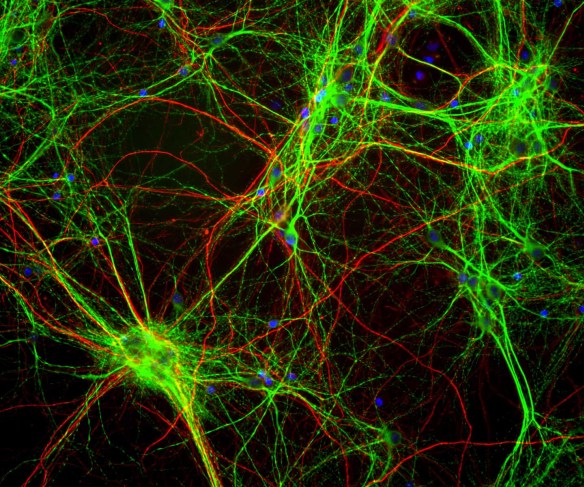
ARG11033 anti-MAP2ab antibody [5H11] ICC/IF image
Immunofluorescence: Mixed neuron/glia cultures stained with ARG11033 anti-MAP2ab antibody [5H11] (green), and co-stained with ARG10761 anti-Neurofilament NF-H antibody (red). DNA stain shows nuclei of neurons and non-neuronal cells (blue).
-
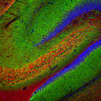
ARG11033 anti-MAP2ab antibody [5H11] IHC-Fr image
Immunohistochemistry: Frozen section of Rat hippocampus stained with ARG11033 anti-MAP2ab antibody [5H11] (green) at 1:5000 dilution and co-stained with ARG10730 anti-NF66 / alpha Internexin antibody (red) at 1:2000 dilution. (Sample preparation: Following transcardial perfusion of Rat with 4% paraformaldehyde, brain was post fixed for 24 hours, cut to 45 µM, and free-floating sections were stained with above antibodies.)
Clone 5H11 labels MAP2 protein in the perikarya and dendrites of the most neurons, and the alpha Internexin antibody selectively stains axons and dendrites of neuronal cells.
-

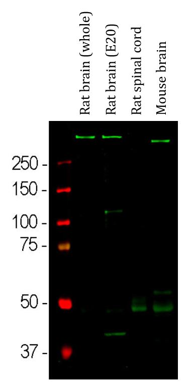
ARG11033 anti-MAP2ab antibody [5H11] WB image
Western blot: Rat brain (whole), Rat brain (embryonic, E20), Rat spinal cord and Mouse brain lysates stained with ARG11033 anti-MAP2ab antibody [5H11] (green) at 1:10000 dilution.
A band at about 280 kDa corresponds to full length MAP2A and MAP2B protein. MAP2A/B is expressed heavily in brain particularly in cortical regions, but is a more minor component of spinal cord.
-
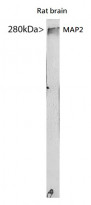
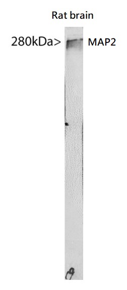
ARG11033 anti-MAP2ab antibody [5H11] WB image
Western blot: Whole Rat brain lysate stained with ARG11033 anti-MAP2ab antibody [5H11].
-
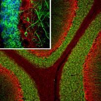
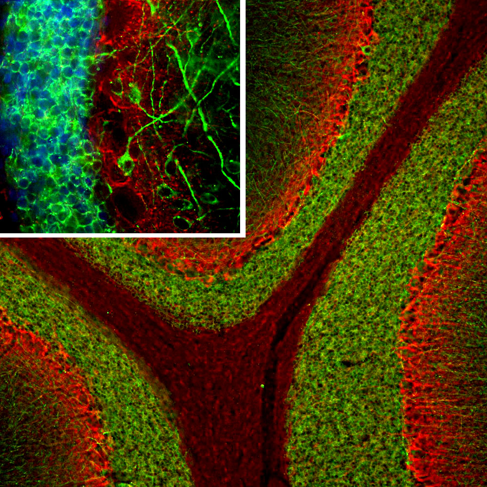
ARG11033 anti-MAP2ab antibody [5H11] IHC-Fr image
Immunohistochemistry: Frozen section of Rat cerebellum stained with ARG11033 anti-MAP2ab antibody [5H11] (green) at 1:5000 dilution and co-stained with ARG10730 anti-NF66 / alpha Internexin antibody (red) at 1:2000 dilution. (Sample preparation: Following transcardial perfusion of Rat with 4% paraformaldehyde, brain was post fixed for 24 hours, cut to 45 µM, and free-floating sections were stained with above antibodies.)
Clone 5H11 labels MAP2 protein in the perikarya and dendrites of the most neurons, particularly in granular cell and molecular layers of the cerebellum, while the alpha Internexin antibody selectively stains axons and dendrites of neuronal cells.