ARG66892
anti-M13 Bacteriophage g8p coat protein antibody [SQab21250]
anti-M13 Bacteriophage g8p coat protein antibody [SQab21250] for ELISA,Flow cytometry and Virus
Overview
| Product Description | Mouse Monoclonal antibody [SQab21250] recognizes M13 Bacteriophage g8p coat protein |
|---|---|
| Tested Reactivity | Virus |
| Tested Application | ELISA, FACS |
| Host | Mouse |
| Clonality | Monoclonal |
| Clone | SQab21250 |
| Isotype | IgG2b |
| Target Name | M13 Bacteriophage g8p coat protein |
| Antigen Species | Virus |
| Immunogen | g8p coat protein combine M13KO7 phage. |
| Conjugation | Un-conjugated |
| Alternate Names | M13 Bacteriophage coat phage protein pVIII; M13 pVIII protein; M13 pVIII coat protein; M13 antibody (g8p); Anti-M13 Bacteriophage g8p Coat Protein Antibody, SQab21250, Arigo Biolaboratories, Western blot, ELISA, immunoprecipitation, M13 bacteriophage, DNA cloning, sequencing, g8p coat protein, molecular biology applications, research use |
Application Instructions
| Application Suggestion |
|
||||||
|---|---|---|---|---|---|---|---|
| Application Note | This antibody reacts phage by ELISA and flow cytometry, but does not reacts to recombinant Fc-tagged g8p protein. * The dilutions indicate recommended starting dilutions and the optimal dilutions or concentrations should be determined by the scientist. |
Properties
| Form | Liquid |
|---|---|
| Purification | Purification with Protein G. |
| Purity | 95% (by SDS-PAGE) |
| Buffer | PBS and 0.01% Sodium azide. |
| Preservative | 0.01% Sodium azide |
| Storage Instruction | For continuous use, store undiluted antibody at 2-8°C for up to a week. For long-term storage, aliquot and store at -20°C or below. Storage in frost free freezers is not recommended. Avoid repeated freeze/thaw cycles. Suggest spin the vial prior to opening. The antibody solution should be gently mixed before use. |
| Note | For laboratory research only, not for drug, diagnostic or other use. |
Bioinformation
| Gene Symbol | VIII (M13) |
|---|---|
| Gene Full Name | Capsid protein G8P (M13) |
| Function | M13 Bacteriophage g8p coat protein self assembles to form a helical capsid wrapping up the viral genomic DNA. The capsid displays a filamentous structure with a length of 760-1950 nm and a width of 6-8 nm. The virion assembly and budding take place at the host inner membrane. [UniProtKB - P69541 (CAPSD_BPM13)] |
| Highlight | Related news: M13 monoclonal antibody validated in ELISA and FACS; |
Images (4) Click the Picture to Zoom In
-
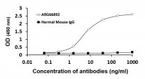
ARG66892 anti-M13 Bacteriophage g8p coat protein antibody [SQab21250] ELISA image
ELISA: M13 phage was pre-coated on the plate and detected the phage by variable concentrations of ARG66892 anti-M13 Bacteriophage g8p coat protein antibody [SQab21250]. Isotype control: Normal mouse IgG.
-
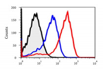
ARG66892 anti-M13 Bacteriophage g8p coat protein antibody [SQab21250] FACS image
Flow Cytometry: MCF7 cells incubated with 1x10^9 (blue) or 1x10^10 (red) of anti-HER3 scFvs displayed phage, then stained with ARG66892 anti-M13 Bacteriophage g8p coat protein antibody [SQab21250] at 1:500 dilution. And a PE-labeled anti-mouse IgG antibody used as the third antibody. Cells did not incubate the anti-HER3 scFvs displayed phage as control sample (black).
-
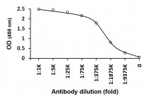
ARG66892 anti-M13 Bacteriophage g8p coat protein antibody [SQab21250] ELISA image
ELISA: The plate was coated with 10^8 of M13 bacteriophages. Samples were detected with serially diluted ARG66892 anti-M13 Bacteriophage g8p coat protein antibody [SQab21250].
-
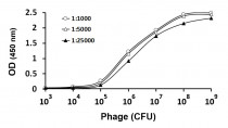
ARG66892 anti-M13 Bacteriophage g8p coat protein antibody [SQab21250] ELISA image
Sensitivity analysis: Coating the indicated amounts of M13 bacteriophages and stained with the different concentration of ARG66892 anti-M13 Bacteriophage g8p coat protein antibody [SQab21250].


