ARG41414
anti-Lyn antibody
anti-Lyn antibody for IHC-Formalin-fixed paraffin-embedded sections and Human,Mouse,Rat
Signaling Transduction antibody; Src Family Protein Tyrosine Kinases antibody
Overview
| Product Description | Rabbit Polyclonal antibody recognizes Lyn |
|---|---|
| Tested Reactivity | Hu, Ms, Rat |
| Tested Application | IHC-P |
| Host | Rabbit |
| Clonality | Polyclonal |
| Isotype | IgG |
| Target Name | Lyn |
| Antigen Species | Human |
| Immunogen | Synthetic peptide corresponding to aa. 470-501 of Human Lyn. (DELYDIMKMCWKEKAEERPTFDYLQSVLDDFY) |
| Conjugation | Un-conjugated |
| Alternate Names | JTK8; Lck/Yes-related novel protein tyrosine kinase; V-yes-1 Yamaguchi sarcoma viral related oncogene homolog; p56Lyn; Tyrosine-protein kinase Lyn; p53Lyn; EC 2.7.10.2 |
Application Instructions
| Application Suggestion |
|
||||
|---|---|---|---|---|---|
| Application Note | IHC-P: Antigen Retrieval: Heat mediation was performed in Citrate buffer (pH 6.0, epitope retrieval solution) for 20 min. * The dilutions indicate recommended starting dilutions and the optimal dilutions or concentrations should be determined by the scientist. |
Properties
| Form | Liquid |
|---|---|
| Purification | Affinity purification with immunogen. |
| Buffer | 0.2% Na2HPO4, 0.9% NaCl, 0.05% Sodium azide and 5% BSA. |
| Preservative | 0.05% Sodium azide |
| Stabilizer | 5% BSA |
| Concentration | 0.5 mg/ml |
| Storage Instruction | For continuous use, store undiluted antibody at 2-8°C for up to a week. For long-term storage, aliquot and store at -20°C or below. Storage in frost free freezers is not recommended. Avoid repeated freeze/thaw cycles. Suggest spin the vial prior to opening. The antibody solution should be gently mixed before use. |
| Note | For laboratory research only, not for drug, diagnostic or other use. |
Bioinformation
| Database Links | |
|---|---|
| Gene Symbol | LYN |
| Gene Full Name | LYN proto-oncogene, Src family tyrosine kinase |
| Background | This gene encodes a tyrosine protein kinase, which maybe involved in the regulation of mast cell degranulation, and erythroid differentiation. Alternatively spliced transcript variants encoding different isoforms have been found for this gene. [provided by RefSeq, Jul 2011] |
| Function | Non-receptor tyrosine-protein kinase that transmits signals from cell surface receptors and plays an important role in the regulation of innate and adaptive immune responses, hematopoiesis, responses to growth factors and cytokines, integrin signaling, but also responses to DNA damage and genotoxic agents. Functions primarily as negative regulator, but can also function as activator, depending on the context. Required for the initiation of the B-cell response, but also for its down-regulation and termination. Plays an important role in the regulation of B-cell differentiation, proliferation, survival and apoptosis, and is important for immune self-tolerance. Acts downstream of several immune receptors, including the B-cell receptor, CD79A, CD79B, CD5, CD19, CD22, FCER1, FCGR2, FCGR1A, TLR2 and TLR4. Plays a role in the inflammatory response to bacterial lipopolysaccharide. Mediates the responses to cytokines and growth factors in hematopoietic progenitors, platelets, erythrocytes, and in mature myeloid cells, such as dendritic cells, neutrophils and eosinophils. Acts downstream of EPOR, KIT, MPL, the chemokine receptor CXCR4, as well as the receptors for IL3, IL5 and CSF2. Plays an important role in integrin signaling. Regulates cell proliferation, survival, differentiation, migration, adhesion, degranulation, and cytokine release. Down-regulates signaling pathways by phosphorylation of immunoreceptor tyrosine-based inhibitory motifs (ITIM), that then serve as binding sites for phosphatases, such as PTPN6/SHP-1, PTPN11/SHP-2 and INPP5D/SHIP-1, that modulate signaling by dephosphorylation of kinases and their substrates. Phosphorylates LIME1 in response to CD22 activation. Phosphorylates BTK, CBL, CD5, CD19, CD72, CD79A, CD79B, CSF2RB, DOK1, HCLS1, LILRB3/PIR-B, MS4A2/FCER1B, PTK2B/PYK2, SYK and TEC. Promotes phosphorylation of SIRPA, PTPN6/SHP-1, PTPN11/SHP-2 and INPP5D/SHIP-1. Mediates phosphorylation of the BCR-ABL fusion protein. Required for rapid phosphorylation of FER in response to FCER1 activation. Mediates KIT phosphorylation. Acts as an effector of EPOR (erythropoietin receptor) in controlling KIT expression and may play a role in erythroid differentiation during the switch between proliferation and maturation. Depending on the context, activates or inhibits several signaling cascades. Regulates phosphatidylinositol 3-kinase activity and AKT1 activation. Regulates activation of the MAP kinase signaling cascade, including activation of MAP2K1/MEK1, MAPK1/ERK2, MAPK3/ERK1, MAPK8/JNK1 and MAPK9/JNK2. Mediates activation of STAT5A and/or STAT5B. Phosphorylates LPXN on 'Tyr-72'. Kinase activity facilitates TLR4-TLR6 heterodimerization and signal initiation. [UniProt] |
| Cellular Localization | Cell membrane. Nucleus. Cytoplasm. Cytoplasm, perinuclear region. Golgi apparatus. Membrane; Lipid-anchor. Note=Accumulates in the nucleus by inhibition of CRM1-mediated nuclear export. Nuclear accumulation is increased by inhibition of its kinase activity. The trafficking from the Golgi apparatus to the plasma membrane occurs in a kinase domain-dependent but kinase activity independent manner and is mediated by exocytic vesicular transport. Detected on plasma membrane lipid rafts. [UniProt] |
| Research Area | Signaling Transduction antibody; Src Family Protein Tyrosine Kinases antibody |
| Calculated MW | 59 kDa |
| PTM | Ubiquitinated by CBL, leading to its degradation. Ubiquitination is SH3-dependent. Autophosphorylated. Phosphorylated on tyrosine residues in response to KIT signaling. Phosphorylation at Tyr-397 is required for optimal activity. Phosphorylation at Tyr-508 inhibits kinase activity. Phosphorylated at Tyr-508 by CSK. Dephosphorylated by PTPRC/CD45. Becomes rapidly phosphorylated upon activation of the B-cell receptor and the immunoglobulin receptor FCGR1A. [UniProt] |
Images (5) Click the Picture to Zoom In
-
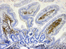
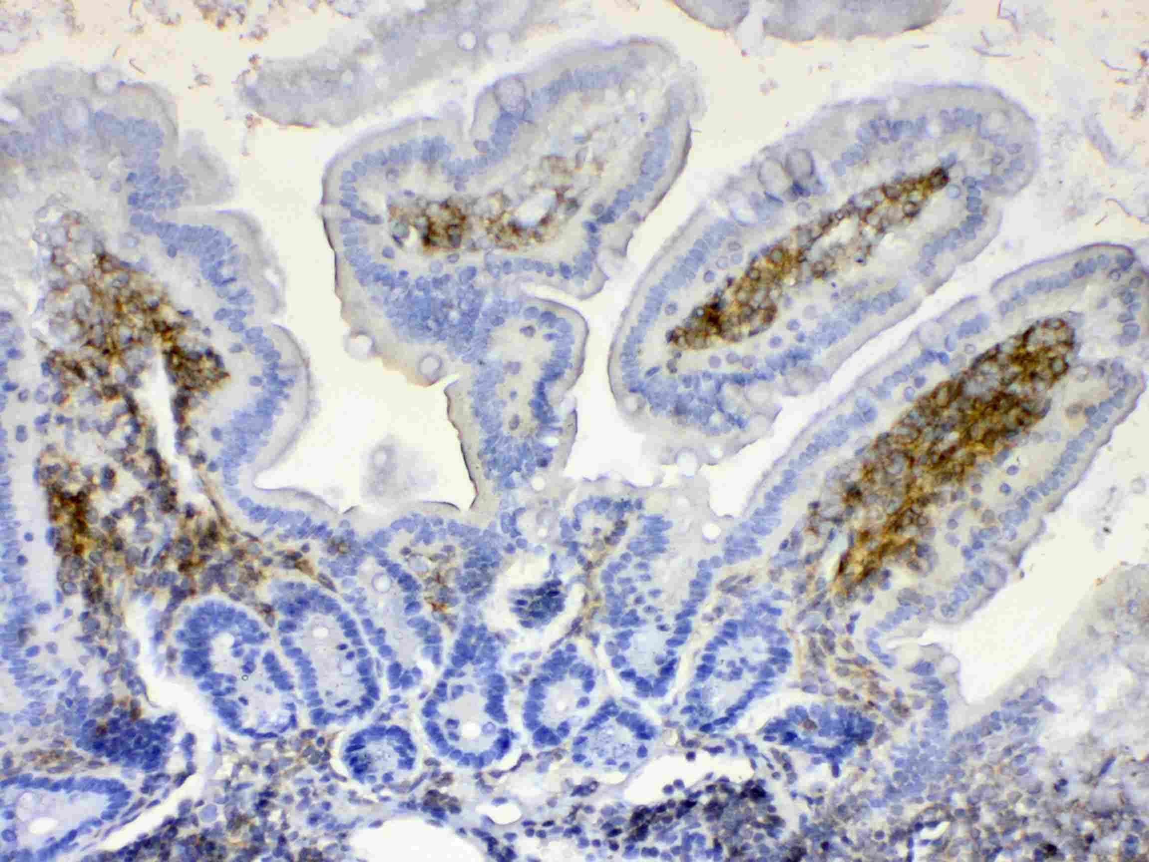
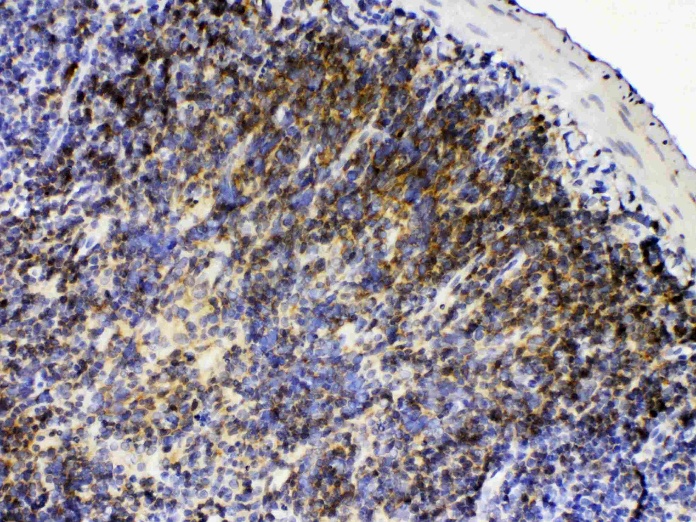
ARG41414 anti-Lyn antibody IHC-P image
Immunohistochemistry: Paraffin-embedded Mouse intestine tissue. Antigen Retrieval: Heat mediation was performed in Citrate buffer (pH 6.0, epitope retrieval solution) for 20 min. The tissue section was blocked with 10% goat serum. The tissue section was then stained with ARG41414 anti-Lyn antibody at 1 µg/ml dilution, overnight at 4°C.
-
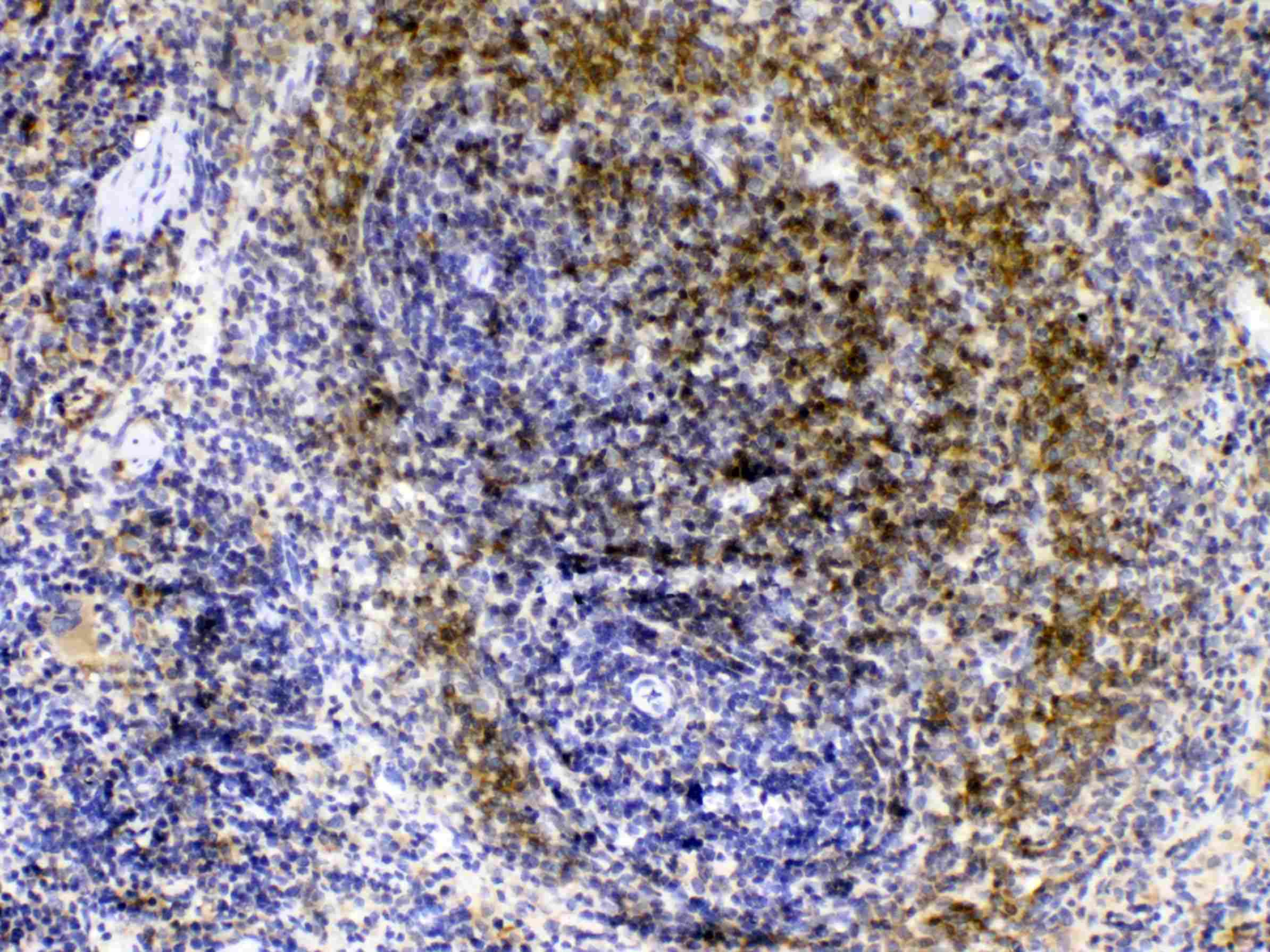
ARG41414 anti-Lyn antibody IHC-P image
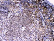
Immunohistochemistry: Paraffin-embedded Rat spleen tissue. Antigen Retrieval: Heat mediation was performed in Citrate buffer (pH 6.0, epitope retrieval solution) for 20 min. The tissue section was blocked with 10% goat serum. The tissue section was then stained with ARG41414 anti-Lyn antibody at 1 µg/ml dilution, overnight at 4°C.
-
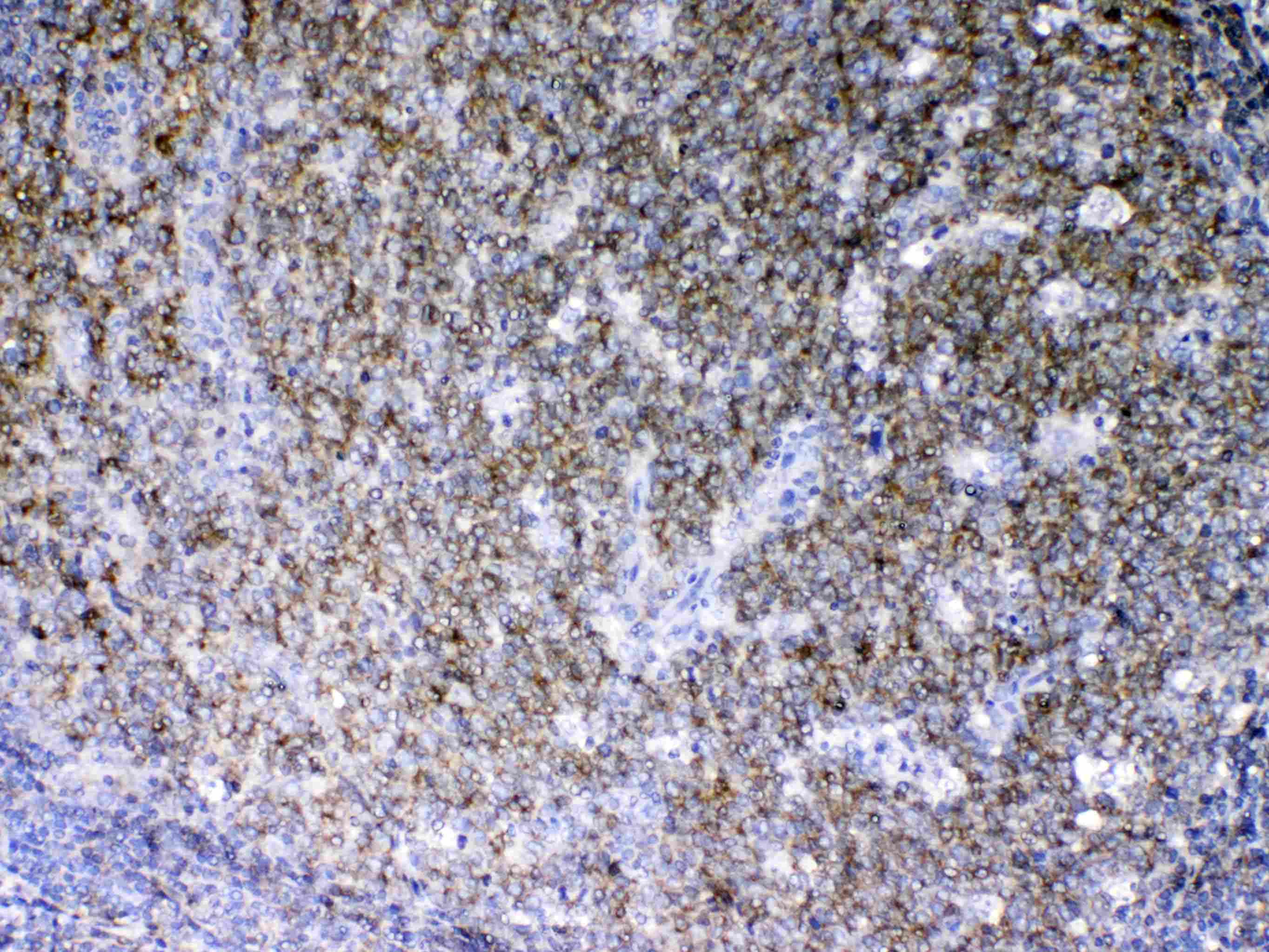
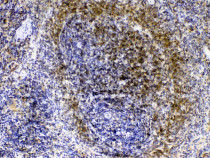
ARG41414 anti-Lyn antibody IHC-P image
Immunohistochemistry: Paraffin-embedded Human tonsil tissue. Antigen Retrieval: Heat mediation was performed in Citrate buffer (pH 6.0, epitope retrieval solution) for 20 min. The tissue section was blocked with 10% goat serum. The tissue section was then stained with ARG41414 anti-Lyn antibody at 1 µg/ml dilution, overnight at 4°C.
-
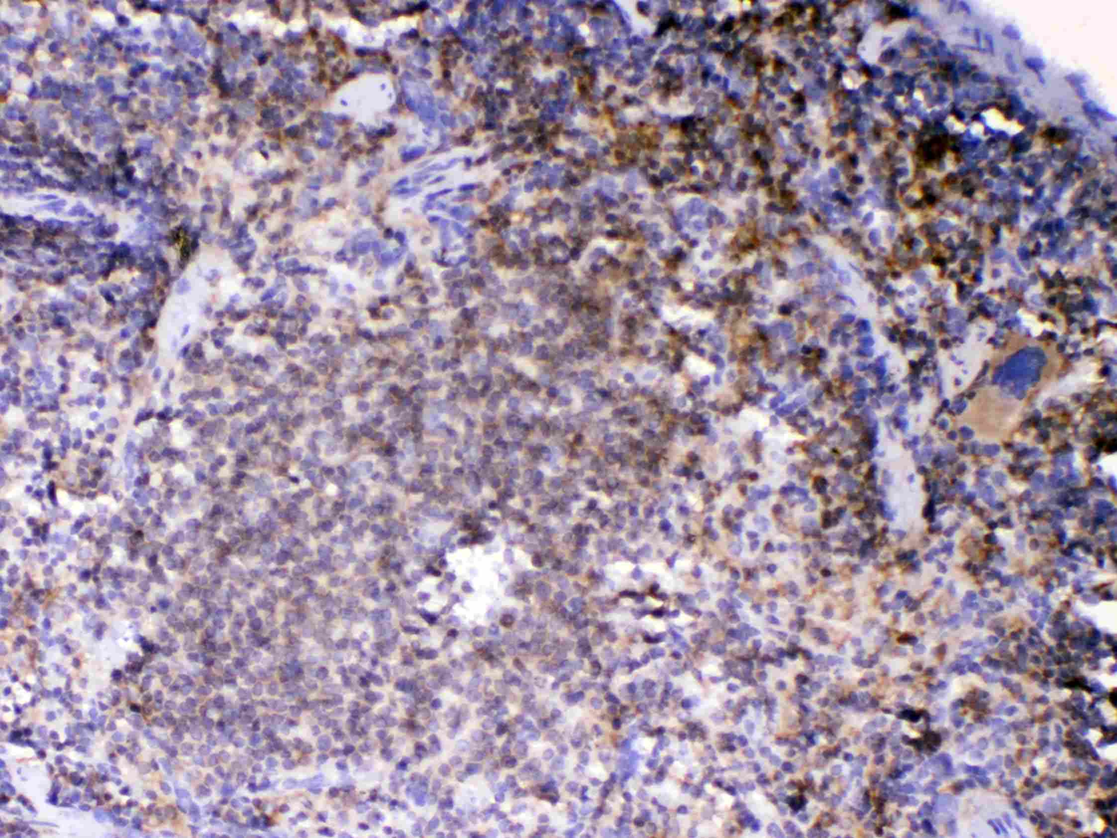
ARG41414 anti-Lyn antibody IHC-P image
Immunohistochemistry: Paraffin-embedded Mouse spleen tissue. Antigen Retrieval: Heat mediation was performed in Citrate buffer (pH 6.0, epitope retrieval solution) for 20 min. The tissue section was blocked with 10% goat serum. The tissue section was then stained with ARG41414 anti-Lyn antibody at 1 µg/ml dilution, overnight at 4°C.
-
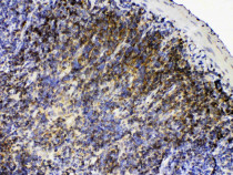
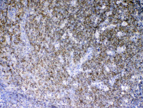
ARG41414 anti-Lyn antibody IHC-P image
Immunohistochemistry: Paraffin-embedded Rat lymphaden tissue. Antigen Retrieval: Heat mediation was performed in Citrate buffer (pH 6.0, epitope retrieval solution) for 20 min. The tissue section was blocked with 10% goat serum. The tissue section was then stained with ARG41414 anti-Lyn antibody at 1 µg/ml dilution, overnight at 4°C.