ARG41423
anti-Lck antibody
anti-Lck antibody for Flow cytometry,ICC/IF,IHC-Frozen sections,IHC-Formalin-fixed paraffin-embedded sections,Western blot and Human,Mouse,Rat
Immune System antibody; Signaling Transduction antibody; Src Family Protein Tyrosine Kinases antibody
Overview
| Product Description | Rabbit Polyclonal antibody recognizes Lck |
|---|---|
| Tested Reactivity | Hu, Ms, Rat |
| Tested Application | FACS, ICC/IF, IHC-Fr, IHC-P, WB |
| Host | Rabbit |
| Clonality | Polyclonal |
| Isotype | IgG |
| Target Name | Lck |
| Antigen Species | Human |
| Immunogen | Synthetic peptide corresponding to aa. 468-506 of Human Lck. (ELYQLMRLCWKERPEDRPTFDYLRSVLEDFFTATEGQYQ) |
| Conjugation | Un-conjugated |
| Alternate Names | T cell-specific protein-tyrosine kinase; Leukocyte C-terminal Src kinase; Protein YT16; p56-LCK; LSK; Proto-oncogene Lck; p56lck; pp58lck; Tyrosine-protein kinase Lck; YT16; IMD22; Lymphocyte cell-specific protein-tyrosine kinase; EC 2.7.10.2 |
Application Instructions
| Application Suggestion |
|
||||||||||||
|---|---|---|---|---|---|---|---|---|---|---|---|---|---|
| Application Note | * The dilutions indicate recommended starting dilutions and the optimal dilutions or concentrations should be determined by the scientist. | ||||||||||||
| Observed Size | ~ 58 kDa |
Properties
| Form | Liquid |
|---|---|
| Purification | Affinity purification with immunogen. |
| Buffer | 0.2% Na2HPO4, 0.9% NaCl, 0.05% Sodium azide and 5% BSA. |
| Preservative | 0.05% Sodium azide |
| Stabilizer | 5% BSA |
| Concentration | 0.5 mg/ml |
| Storage Instruction | For continuous use, store undiluted antibody at 2-8°C for up to a week. For long-term storage, aliquot and store at -20°C or below. Storage in frost free freezers is not recommended. Avoid repeated freeze/thaw cycles. Suggest spin the vial prior to opening. The antibody solution should be gently mixed before use. |
| Note | For laboratory research only, not for drug, diagnostic or other use. |
Bioinformation
| Database Links | |
|---|---|
| Gene Symbol | LCK |
| Gene Full Name | LCK proto-oncogene, Src family tyrosine kinase |
| Background | This gene is a member of the Src family of protein tyrosine kinases (PTKs). The encoded protein is a key signaling molecule in the selection and maturation of developing T-cells. It contains N-terminal sites for myristylation and palmitylation, a PTK domain, and SH2 and SH3 domains which are involved in mediating protein-protein interactions with phosphotyrosine-containing and proline-rich motifs, respectively. The protein localizes to the plasma membrane and pericentrosomal vesicles, and binds to cell surface receptors, including CD4 and CD8, and other signaling molecules. Multiple alternatively spliced variants, encoding the same protein, have been described. [provided by RefSeq, Jul 2008] |
| Function | Non-receptor tyrosine-protein kinase that plays an essential role in the selection and maturation of developing T-cells in the thymus and in the function of mature T-cells. Plays a key role in T-cell antigen receptor (TCR)-linked signal transduction pathways. Constitutively associated with the cytoplasmic portions of the CD4 and CD8 surface receptors. Association of the TCR with a peptide antigen-bound MHC complex facilitates the interaction of CD4 and CD8 with MHC class II and class I molecules, respectively, thereby recruiting the associated LCK protein to the vicinity of the TCR/CD3 complex. LCK then phosphorylates tyrosines residues within the immunoreceptor tyrosine-based activation motifs (ITAM) of the cytoplasmic tails of the TCR-gamma chains and CD3 subunits, initiating the TCR/CD3 signaling pathway. Once stimulated, the TCR recruits the tyrosine kinase ZAP70, that becomes phosphorylated and activated by LCK. Following this, a large number of signaling molecules are recruited, ultimately leading to lymphokine production. LCK also contributes to signaling by other receptor molecules. Associates directly with the cytoplasmic tail of CD2, which leads to hyperphosphorylation and activation of LCK. Also plays a role in the IL2 receptor-linked signaling pathway that controls the T-cell proliferative response. Binding of IL2 to its receptor results in increased activity of LCK. Is expressed at all stages of thymocyte development and is required for the regulation of maturation events that are governed by both pre-TCR and mature alpha beta TCR. Phosphorylates other substrates including RUNX3, PTK2B/PYK2, the microtubule-associated protein MAPT, RHOH or TYROBP. [UniProt] |
| Cellular Localization | Cytoplasm. Cell membrane; Lipid-anchor; Cytoplasmic side. Note=Present in lipid rafts in an inactive form. [UniProt] |
| Research Area | Immune System antibody; Signaling Transduction antibody; Src Family Protein Tyrosine Kinases antibody |
| Calculated MW | 58 kDa |
| PTM | Autophosphorylated on Tyr-394, increasing enzymatic activity, this site is dephosphorylated by PTN22. Phosphorylated on Tyr-505 by CSK, decreasing activity. Dephosphorylated by PTPRC/CD45. Dephosphorylation at Tyr-394 by PTPN2 negatively regulates T-cell receptor signaling. Myristoylation is required prior to palmitoylation. Palmitoylation regulates subcellular location. [UniProt] |
Images (7) Click the Picture to Zoom In
-
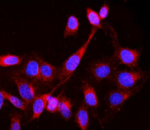
ARG41423 anti-Lck antibody ICC/IF image
Immunofluorescence: U2OS cells were blocked with 10% goat serum and then stained with ARG41423 anti-Lck antibody (red) at 2 µg/ml dilution, overnight at 4°C. DAPI (blue) for nuclear staining.
-

ARG41423 anti-Lck antibody IHC-Fr image
Immunohistochemistry: Frozen section of Mouse spleen tissue. The tissue section was blocked with 10% goat serum. The tissue section was then stained with ARG41423 anti-Lck antibody at 1 µg/ml dilution, overnight at 4°C.
-
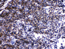
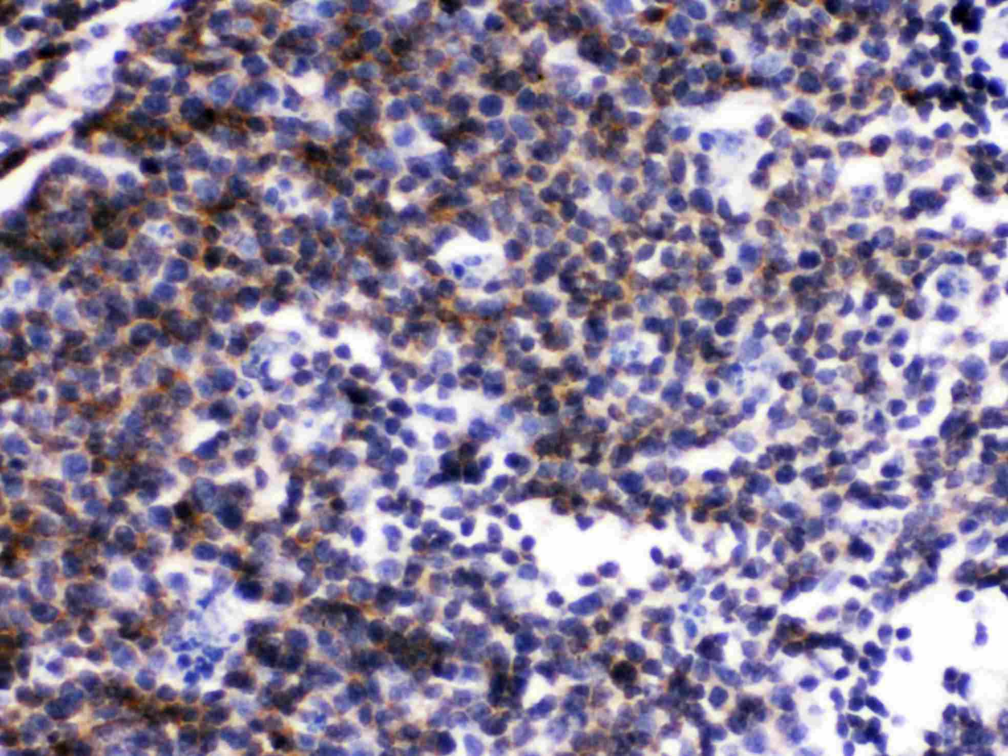
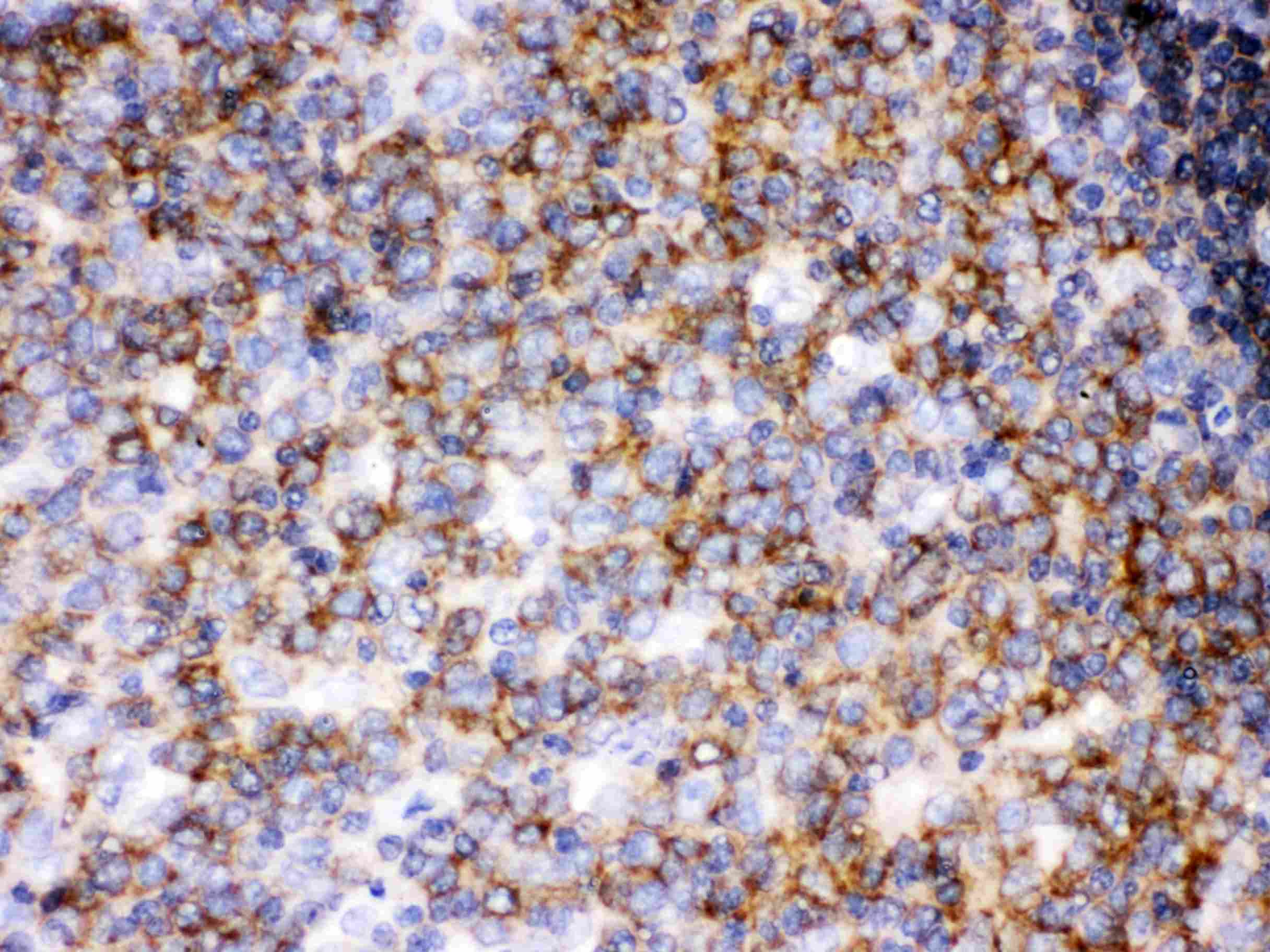
ARG41423 anti-Lck antibody IHC-P image
Immunohistochemistry: Paraffin-embedded Mouse lymphaden tissue stained with ARG41423 anti-Lck antibody.
-
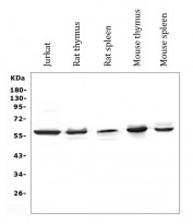
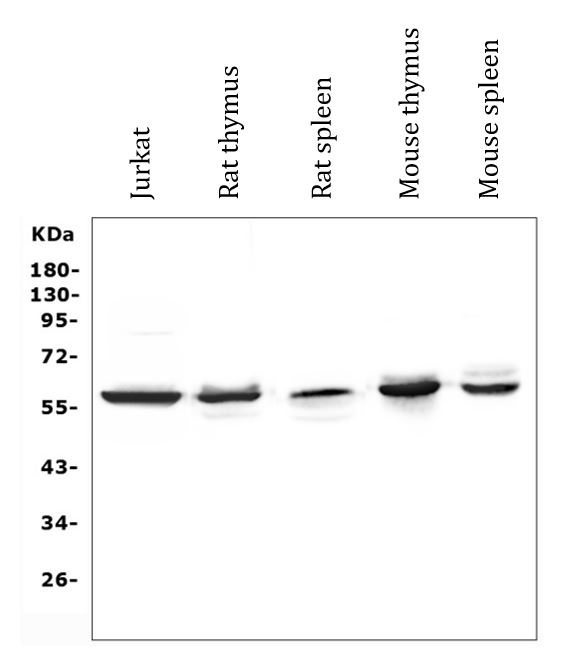
ARG41423 anti-Lck antibody WB image
Western blot: 50 µg of samples under reducing conditions. Jurkat, Rat thymus, Rat spleen, Mouse thymus and Mouse spleen lysates stained with ARG41423 anti-Lck antibody at 0.5 µg/ml dilution, overnight at 4°C.
-
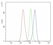
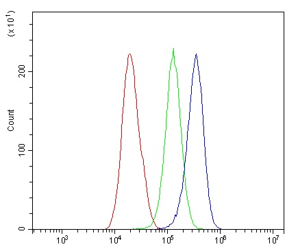
ARG41423 anti-Lck antibody FACS image
Flow Cytometry: HepG2 cells were blocked with 10% normal goat serum and then stained with ARG41423 anti-Lck antibody (blue) at 1 µg/10^6 cells for 30 min at 20°C, followed by incubation with DyLight®488 labelled secondary antibody. Isotype control antibody (green) was rabbit IgG (1 µg/10^6 cells) used under the same conditions. Unlabelled sample (red) was also used as a control.
-
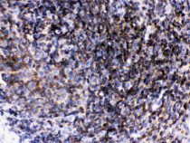
ARG41423 anti-Lck antibody IHC-P image
Immunohistochemistry: Paraffin-embedded Rat lymphaden tissue stained with ARG41423 anti-Lck antibody.
-

ARG41423 anti-Lck antibody IHC-P image
Immunohistochemistry: Paraffin-embedded Human tonsil tissue stained with ARG41423 anti-Lck antibody.