ARG44241
anti-Langerin / CD207 antibody [2G3]
anti-Langerin / CD207 antibody [2G3] for ELISA,Flow cytometry,ICC/IF,IHC-Frozen sections and Human,Primates
Overview
| Product Description | Mouse Monoclonal antibody [2G3] recognizes Langerin / CD207 |
|---|---|
| Tested Reactivity | Hu, NHuPrm |
| Tested Application | ELISA, FACS, ICC/IF, IHC-Fr |
| Host | Mouse |
| Clonality | Monoclonal |
| Clone | 2G3 |
| Isotype | IgG1, lambda |
| Target Name | Langerin / CD207 |
| Antigen Species | Human |
| Immunogen | Human CD207 Fusion protein |
| Conjugation | Un-conjugated |
| Alternate Names | CD207; CD207 Molecule; CLEC4K; C-Type Lectin Domain Family 4 Member K Langerin; Langerhans Cell Specific C-Type Lectin; CD207 Molecule, Langerin; CD207 Antigen, Langerin; C-Type Lectin Domain Family 4, Member K; CD207 Antigen |
Application Instructions
| Application Suggestion |
|
||||||||||
|---|---|---|---|---|---|---|---|---|---|---|---|
| Application Note | * The dilutions indicate recommended starting dilutions and the optimal dilutions or concentrations should be determined by the scientist. |
Properties
| Form | Liquid |
|---|---|
| Purification | Purification with Protein A. |
| Buffer | PBS (pH 7.4) and 15 mM Sodium azide. |
| Preservative | 15 mM Sodium azide |
| Concentration | 1 mg/ml |
| Storage Instruction | Aliquot and store in the dark at 4°C. Keep protected from prolonged exposure to light. Do not freeze. Suggest spin the vial prior to opening. The antibody solution should be gently mixed before use. |
| Note | For laboratory research only, not for drug, diagnostic or other use. |
Bioinformation
| Database Links |
Swiss-port # Q9UJ71 Human C-type lectin domain family 4 member K |
|---|---|
| Gene Symbol | CD207 |
| Gene Full Name | CD207 Molecule |
| Background | The protein encoded by this gene is expressed only in Langerhans cells which are immature dendritic cells of the epidermis and mucosa. It is localized in the Birbeck granules, organelles present in the cytoplasm of Langerhans cells and consisting of superimposed and zippered membranes. It is a C-type lectin with mannose binding specificity, and it has been proposed that mannose binding by this protein leads to internalization of antigen into Birbeck granules and providing access to a nonclassical antigen-processing pathway. Mutations in this gene result in Birbeck granules deficiency or loss of sugar binding activity. |
| Function | Calcium-dependent lectin displaying mannose-binding specificity. Induces the formation of Birbeck granules (BGs); is a potent regulator of membrane superimposition and zippering. Binds to sulfated as well as mannosylated glycans, keratan sulfate (KS) and beta-glucans. Facilitates uptake of antigens and is involved in the routing and/or processing of antigen for presentation to T cells. Major receptor on primary Langerhans cells for Candida species, Saccharomyces species, and Malassezia furfur. Protects against human immunodeficiency virus-1 (HIV-1) infection. Binds to high-mannose structures present on the envelope glycoprotein which is followed by subsequent targeting of the virus to the Birbeck granules leading to its rapid degradation. |
| Cellular Localization | Membrane |
| PTM | Disulfide bond, Glycoprotein |
Images (1) Click the Picture to Zoom In
-

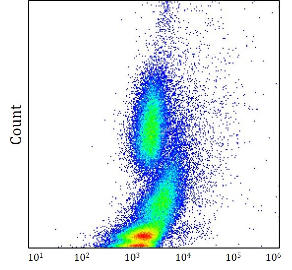
ARG44241 anti-Langerin / CD207 antibody [2G3] FACS image
Flow Cytometry: Human whole blood stained with ARG44241 anti-Langerin / CD207 antibody [2G3] at 0.56 μg/ml dilution.