ARG59198
anti-Laminin antibody
anti-Laminin antibody for IHC-Formalin-fixed paraffin-embedded sections,Western blot and Human,Mouse,Rat
Overview
| Product Description | Rabbit Polyclonal antibody recognizes Laminin |
|---|---|
| Tested Reactivity | Hu, Ms, Rat |
| Tested Application | IHC-P, WB |
| Specificity | The antibody reacts to Laminin gamma 1, 2 and 3. |
| Host | Rabbit |
| Clonality | Polyclonal |
| Isotype | IgG |
| Target Name | Laminin |
| Antigen Species | Human |
| Immunogen | Peptide mixture of laminin gamma 1, 2, 3 (NKLNEIEGSLNKAKDEMKAS; DLEERVRRQRNHLHLLETSI; LQLDSHGALHHKLRQLEEES). |
| Conjugation | Un-conjugated |
| Alternate Names | Laminin subunit gamma-1; Laminin-6 subunit gamma; S-LAM gamma; Laminin-7 subunit gamma; Laminin-2 subunit gamma; Laminin-8 subunit gamma; Laminin-3 subunit gamma; Laminin-10 subunit gamma; Laminin-1 subunit gamma; S-laminin subunit gamma; Laminin B2 chain; Laminin-9 subunit gamma; Laminin-11 subunit gamma; LAMB2; Laminin-4 subunit gamma |
Application Instructions
| Application Suggestion |
|
||||||
|---|---|---|---|---|---|---|---|
| Application Note | IHC-P: Antigen Retrieval: Heat mediated was performed in Citrate buffer (pH 6.0) for 20 min. * The dilutions indicate recommended starting dilutions and the optimal dilutions or concentrations should be determined by the scientist. |
Properties
| Form | Liquid |
|---|---|
| Purification | Affinity purification with immunogen. |
| Buffer | 0.9% NaCl, 0.2% Na2HPO4, 0.05% Sodium azide and 5% BSA. |
| Preservative | 0.05% Sodium azide |
| Stabilizer | 5% BSA |
| Concentration | 0.5 mg/ml |
| Storage Instruction | For continuous use, store undiluted antibody at 2-8°C for up to a week. For long-term storage, aliquot and store at -20°C or below. Storage in frost free freezers is not recommended. Avoid repeated freeze/thaw cycles. Suggest spin the vial prior to opening. The antibody solution should be gently mixed before use. |
| Note | For laboratory research only, not for drug, diagnostic or other use. |
Bioinformation
| Database Links | |
|---|---|
| Gene Symbol | LAMC1 |
| Gene Full Name | laminin, gamma 1 (formerly LAMB2) |
| Background | Laminins, a family of extracellular matrix glycoproteins, are the major noncollagenous constituent of basement membranes. They have been implicated in a wide variety of biological processes including cell adhesion, differentiation, migration, signaling, neurite outgrowth and metastasis. Laminins, composed of 3 non identical chains: laminin alpha, beta and gamma (formerly A, B1, and B2, respectively), have a cruciform structure consisting of 3 short arms, each formed by a different chain, and a long arm composed of all 3 chains. Each laminin chain is a multidomain protein encoded by a distinct gene. Several isoforms of each chain have been described. Different alpha, beta and gamma chain isomers combine to give rise to different heterotrimeric laminin isoforms which are designated by Arabic numerals in the order of their discovery, i.e. alpha1beta1gamma1 heterotrimer is laminin 1. The biological functions of the different chains and trimer molecules are largely unknown, but some of the chains have been shown to differ with respect to their tissue distribution, presumably reflecting diverse functions in vivo. This gene encodes the gamma chain isoform laminin, gamma 1. The gamma 1 chain, formerly thought to be a beta chain, contains structural domains similar to beta chains, however, lacks the short alpha region separating domains I and II. The structural organization of this gene also suggested that it had diverged considerably from the beta chain genes. Embryos of transgenic mice in which both alleles of the gamma 1 chain gene were inactivated by homologous recombination, lacked basement membranes, indicating that laminin, gamma 1 chain is necessary for laminin heterotrimer assembly. It has been inferred by analogy with the strikingly similar 3' UTR sequence in mouse laminin gamma 1 cDNA, that multiple polyadenylation sites are utilized in human to generate the 2 different sized mRNAs (5.5 and 7.5 kb) seen on Northern analysis. [provided by RefSeq, Aug 2011] |
| Function | Binding to cells via a high affinity receptor, laminin is thought to mediate the attachment, migration and organization of cells into tissues during embryonic development by interacting with other extracellular matrix components. [UniProt] |
| Cellular Localization | Secreted, extracellular space, extracellular matrix, basement membrane. [UniProt] |
| Calculated MW | 178 kDa |
Images (5) Click the Picture to Zoom In
-
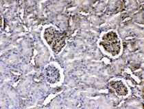
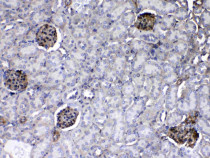
ARG59198 anti-Laminin antibody IHC-P image
Immunohistochemistry: Paraffin-embedded Rat kidney tissue. Antigen Retrieval: Heat mediated was performed in Citrate buffer (pH 6.0, epitope retrieval solution) for 20 min. The tissue section was blocked with 10% goat serum. The tissue section was then stained with ARG59198 anti-Laminin antibody at 1 µg/ml dilution, overnight at 4°C.
-
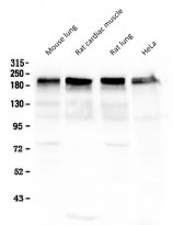
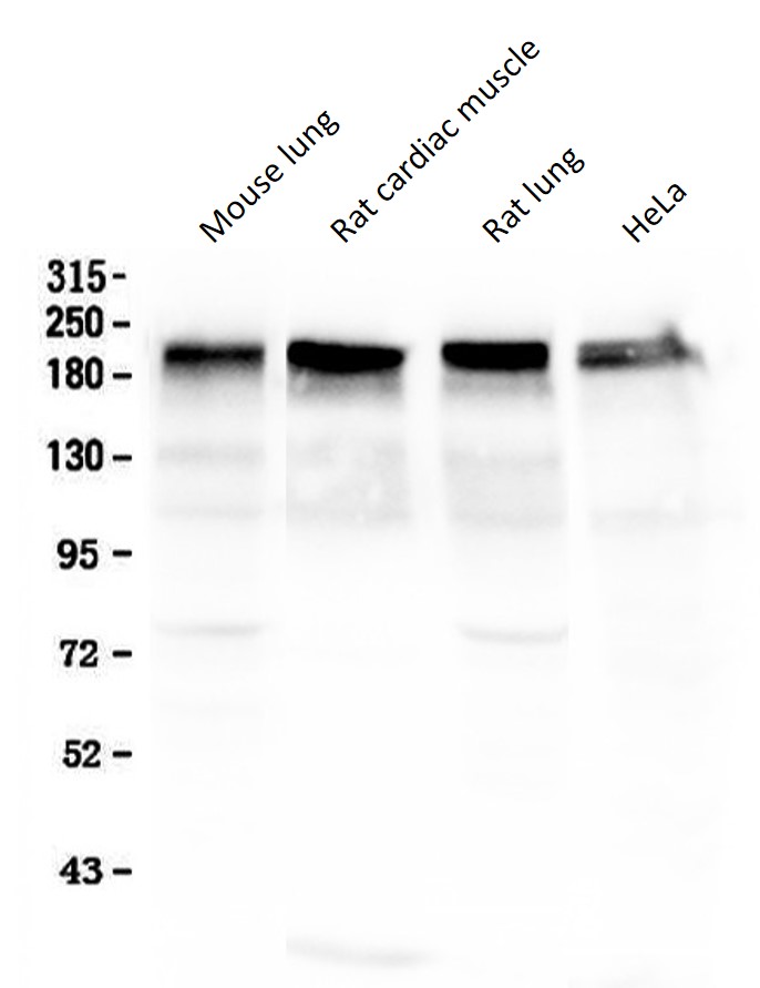
ARG59198 anti-Laminin antibody WB image
Western blot: 50 µg of samples under reducing conditions. Mouse lung, Rat cardiac muscle, Rat lung and HeLa whole cell lysate stained with ARG59198 anti-Laminin antibody at 0.5 µg/ml, overnight at 4°C.
-
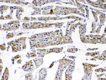
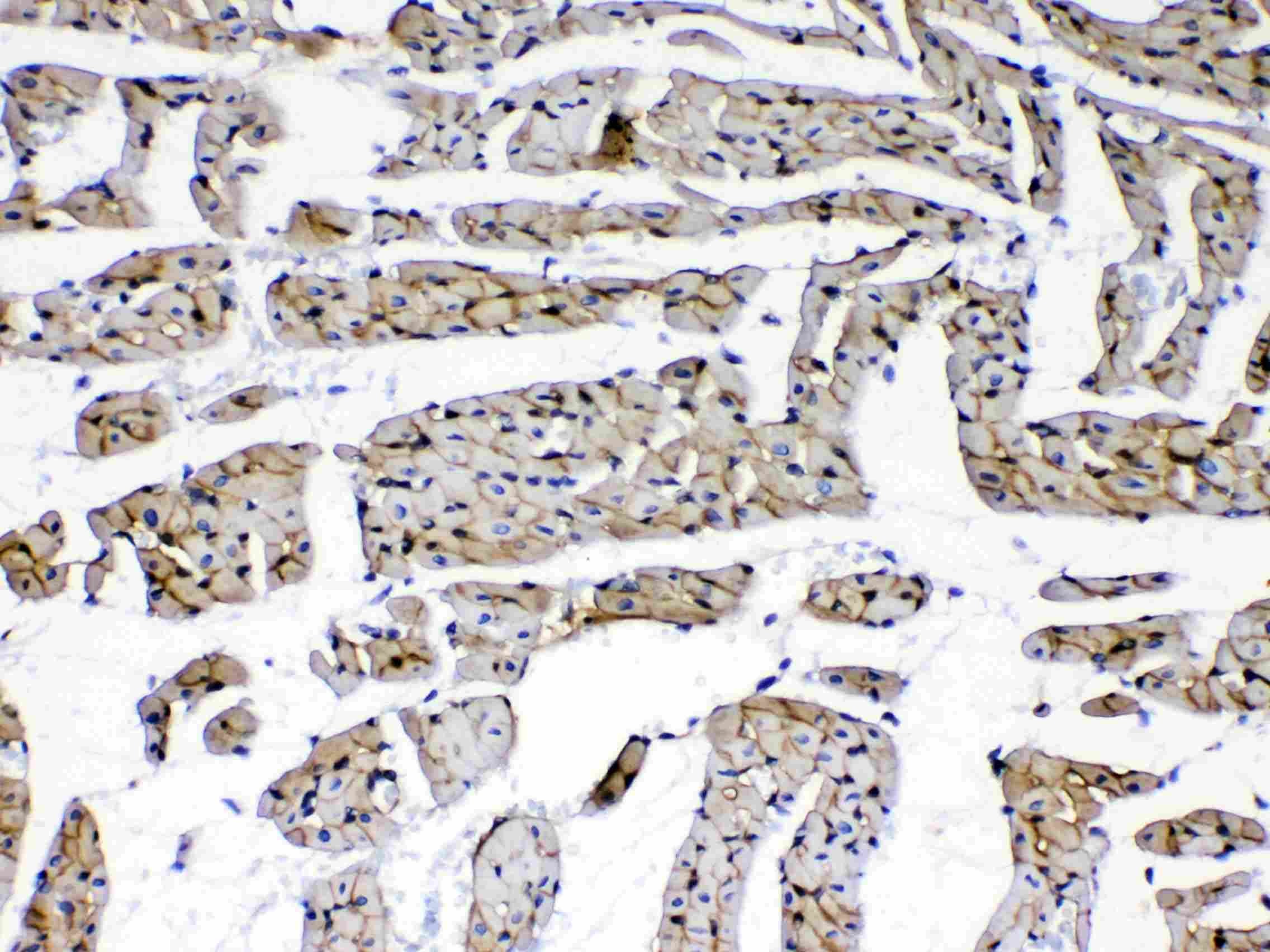
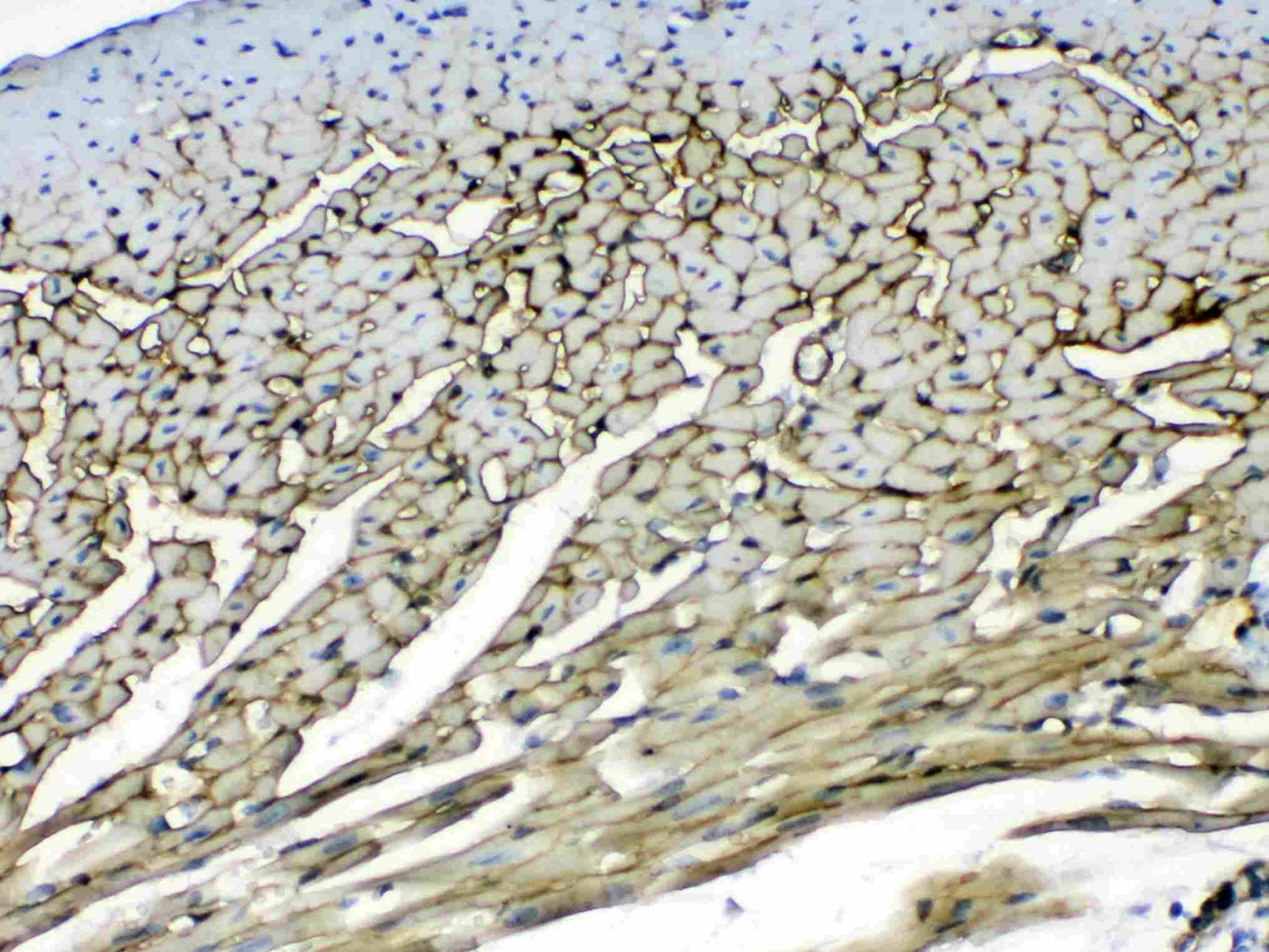
ARG59198 anti-Laminin antibody IHC-P image
Immunohistochemistry: Paraffin-embedded Rat cardiac muscle tissue. Antigen Retrieval: Heat mediated was performed in Citrate buffer (pH 6.0, epitope retrieval solution) for 20 min. The tissue section was blocked with 10% goat serum. The tissue section was then stained with ARG59198 anti-Laminin antibody at 1 µg/ml dilution, overnight at 4°C.
-
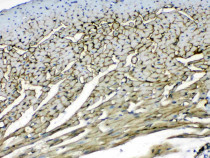
ARG59198 anti-Laminin antibody IHC-P image
Immunohistochemistry: Paraffin-embedded Mouse heart tissue. Antigen Retrieval: Heat mediated was performed in Citrate buffer (pH 6.0, epitope retrieval solution) for 20 min. The tissue section was blocked with 10% goat serum. The tissue section was then stained with ARG59198 anti-Laminin antibody at 1 µg/ml dilution, overnight at 4°C.
-
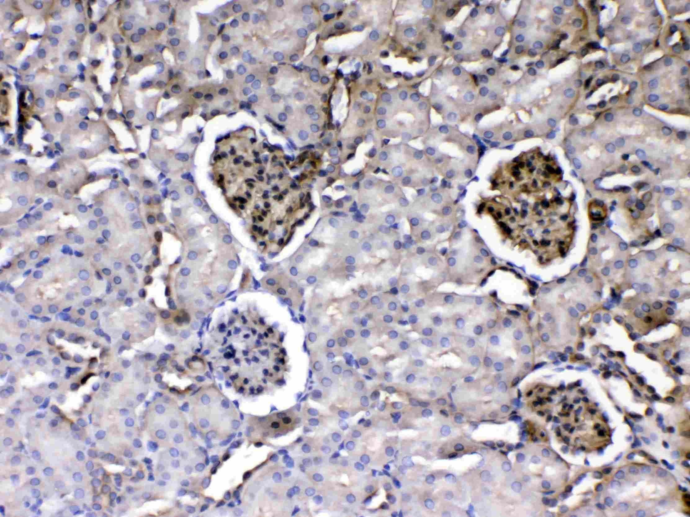
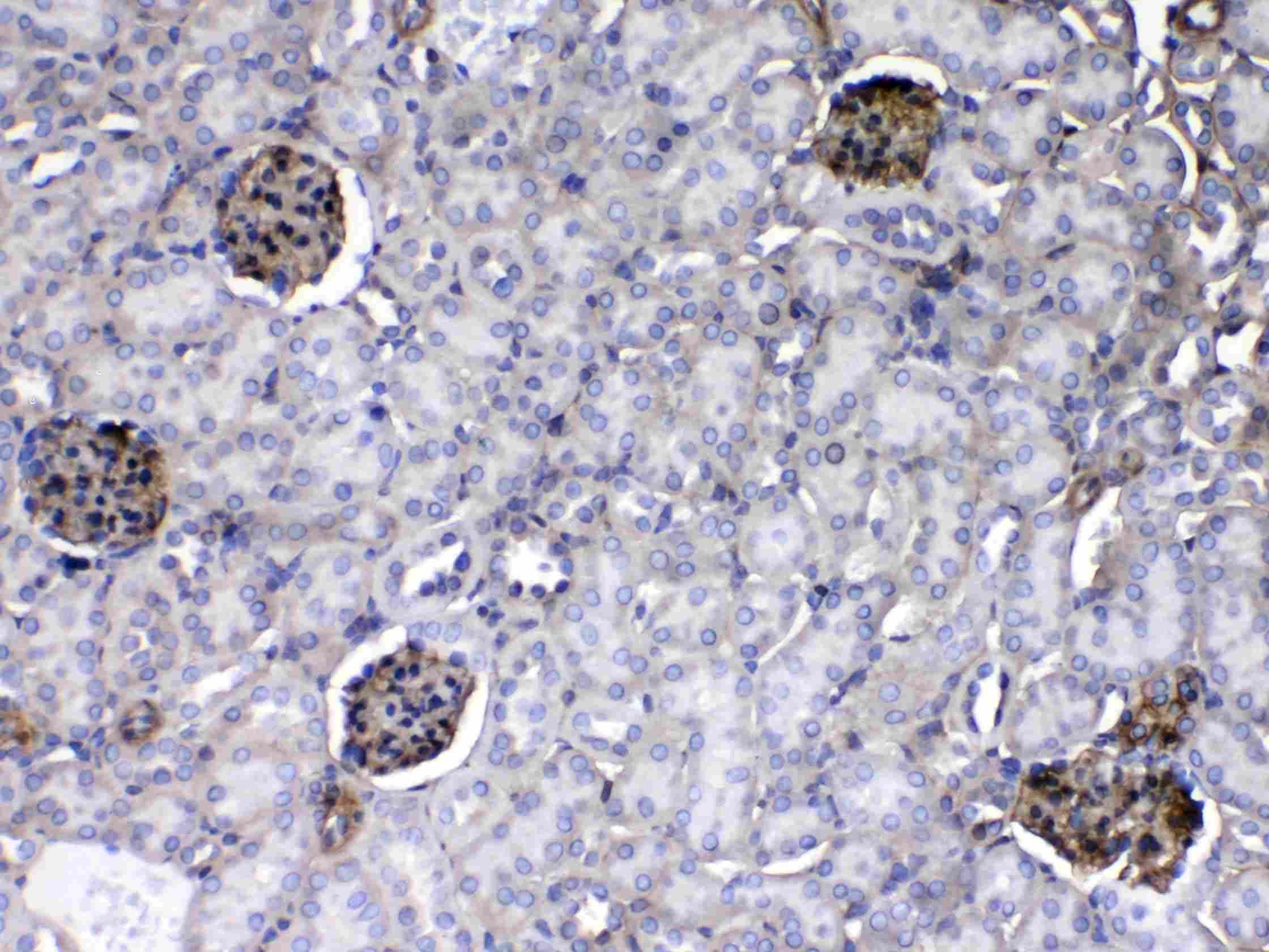
ARG59198 anti-Laminin antibody IHC-P image
Immunohistochemistry: Paraffin-embedded Mouse kidney tissue. Antigen Retrieval: Heat mediated was performed in Citrate buffer (pH 6.0, epitope retrieval solution) for 20 min. The tissue section was blocked with 10% goat serum. The tissue section was then stained with ARG59198 anti-Laminin antibody at 1 µg/ml dilution, overnight at 4°C.
Specific References
Apelin/APJ alleviates diabetic nephropathy by improving glomerular endothelial cells dysfunction via SIRT3‑KLF15
IHC-P / Mouse


Anticancer Effects and Molecular Mechanisms of Apigenin in Cervical Cancer Cells
WB, IHC-P / Human


