ARG41972
anti-Lamin B Receptor antibody
anti-Lamin B Receptor antibody for Flow cytometry,ICC/IF,IHC-Formalin-fixed paraffin-embedded sections,Western blot and Human,Mouse,Rat
Overview
| Product Description | Rabbit Polyclonal antibody recognizes Lamin B Receptor |
|---|---|
| Tested Reactivity | Hu, Ms, Rat |
| Tested Application | FACS, ICC/IF, IHC-P, WB |
| Host | Rabbit |
| Clonality | Polyclonal |
| Isotype | IgG |
| Target Name | Lamin B Receptor |
| Antigen Species | Human |
| Immunogen | Recombinant protein corresponding to H102-F209 of Human Lamin B Receptor. |
| Conjugation | Un-conjugated |
| Alternate Names | PHA; LMN2R; TDRD18; DHCR14B; Integral nuclear envelope inner membrane protein; Lamin-B receptor |
Application Instructions
| Application Suggestion |
|
||||||||||
|---|---|---|---|---|---|---|---|---|---|---|---|
| Application Note | IHC-P: Antigen Retrieval: Heat mediation was performed in Citrate buffer (pH 6.0) for 20 min. * The dilutions indicate recommended starting dilutions and the optimal dilutions or concentrations should be determined by the scientist. |
||||||||||
| Observed Size | ~ 70 kDa |
Properties
| Form | Liquid |
|---|---|
| Purification | Affinity purification with immunogen. |
| Buffer | 0.2% Na2HPO4, 0.9% NaCl, 0.05% Sodium azide and 4% Trehalose. |
| Preservative | 0.05% Sodium azide |
| Stabilizer | 4% Trehalose |
| Concentration | 0.5 mg/ml |
| Storage Instruction | For continuous use, store undiluted antibody at 2-8°C for up to a week. For long-term storage, aliquot and store at -20°C or below. Storage in frost free freezers is not recommended. Avoid repeated freeze/thaw cycles. Suggest spin the vial prior to opening. The antibody solution should be gently mixed before use. |
| Note | For laboratory research only, not for drug, diagnostic or other use. |
Bioinformation
| Database Links | |
|---|---|
| Gene Symbol | LBR |
| Gene Full Name | lamin B receptor |
| Background | The protein encoded by this gene belongs to the ERG4/ERG24 family. It localized in the nuclear envelope inner membrane and anchors the lamina and the heterochromatin to the membrane. It may mediate interaction between chromatin and lamin B. Mutations of this gene has been associated with autosomal recessive HEM/Greenberg skeletal dysplasia. Alternative splicing occurs at this locus and two transcript variants encoding the same protein have been identified. [provided by RefSeq, Jul 2008] |
| Function | Anchors the lamina and the heterochromatin to the inner nuclear membrane. [UniProt] |
| Cellular Localization | Nucleus inner membrane; Multi-pass membrane protein. [UniProt] |
| Calculated MW | 71 kDa |
| PTM | Phosphorylated by CDK1 in mitosis when the inner nuclear membrane breaks down into vesicles that dissociate from the lamina and the chromatin. It is phosphorylated by different protein kinases in interphase when the membrane is associated with these structures. Phosphorylation of LBR and HP1 proteins may be responsible for some of the alterations in chromatin organization and nuclear structure which occur at various times during the cell cycle. Phosphorylated by SRPK1. In late anaphase LBR is dephosphorylated, probably by PP1 and/or PP2A, allowing reassociation with chromatin. [UniProt] |
Images (5) Click the Picture to Zoom In
-
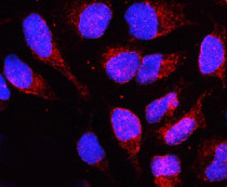
ARG41972 anti-Lamin B Receptor antibody ICC/IF image
Immunofluorescence: U2OS cells were blocked with 10% goat serum and then stained with ARG41972 anti-Lamin B Receptor antibody (red) at 2 µg/ml dilution, overnight at 4°C. DAPI (blue) for nuclear staining.
-
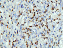
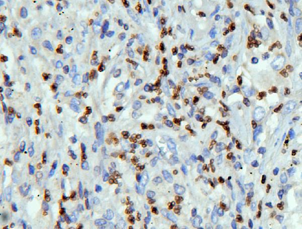
ARG41972 anti-Lamin B Receptor antibody IHC-P image
Immunohistochemistry: Paraffin-embedded Human lung cancer tissue. Antigen Retrieval: Heat mediation was performed in Citrate buffer (pH 6.0, epitope retrieval solution) for 20 min. The tissue section was blocked with 10% goat serum. The tissue section was then stained with ARG41972 anti-Lamin B Receptor antibody at 1 µg/ml dilution, overnight at 4°C.
-

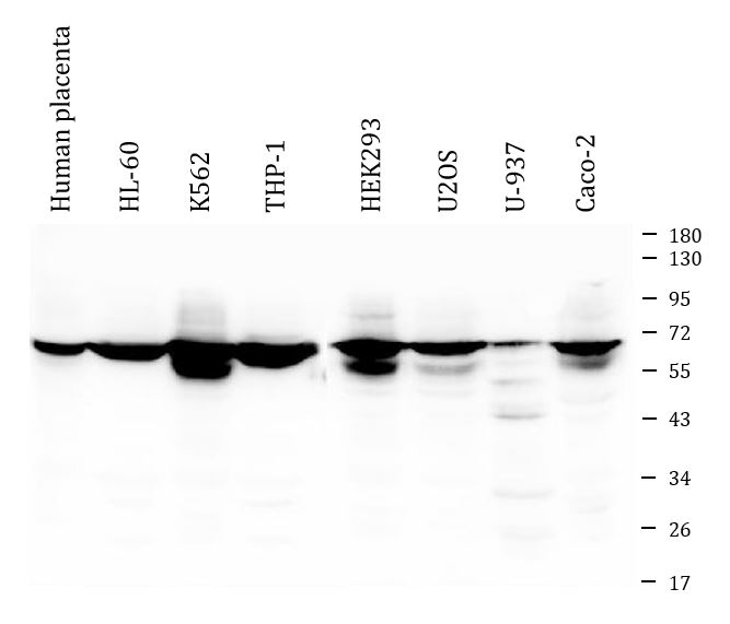
ARG41972 anti-Lamin B Receptor antibody WB image
Western blot: 50 µg of samples under reducing conditions. Human placenta, HL-60, K562, THP-1, HEK293, U2OS, U-937 and Caco-2 whole cell lysates stained with ARG41972 anti-Lamin B Receptor antibody at 0.5 µg/ml, overnight at 4°C.
-
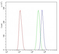
ARG41972 anti-Lamin B Receptor antibody FACS image
Flow Cytometry: U2OS cells were blocked with 10% normal goat serum and then stained with ARG41972 anti-Lamin B Receptor antibody (blue) at 1 µg/10^6 cells for 30 min at 20°C, followed by incubation with DyLight®488 labelled secondary antibody. Isotype control antibody (green) was rabbit IgG (1 µg/10^6 cells) used under the same conditions. Unlabelled sample (red) was also used as a control.
-
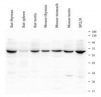
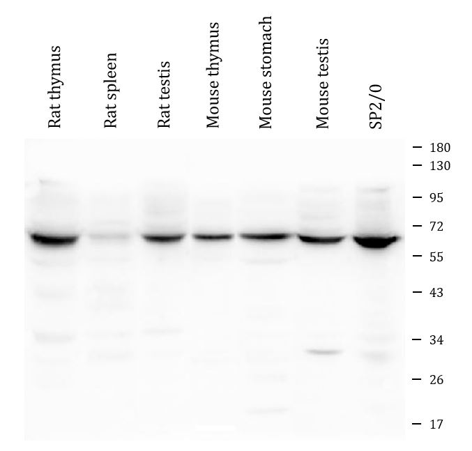
ARG41972 anti-Lamin B Receptor antibody WB image
Western blot: 50 µg of samples under reducing conditions. Rat thymus, Rat spleen, Rat testis, Mouse thymus, Mouse stomach, Mouse testis and Mouse SP2/0 whole cell lysates stained with ARG41972 anti-Lamin B Receptor antibody at 0.5 µg/ml dilution, overnight at 4°C.