ARG42015
anti-Lamin A + C antibody
anti-Lamin A + C antibody for ICC/IF,IHC-Formalin-fixed paraffin-embedded sections,Immunoprecipitation,Western blot and Human,Mouse,Rat
Overview
| Product Description | Rabbit Polyclonal antibody recognizes Lamin A + C |
|---|---|
| Tested Reactivity | Hu, Ms, Rat |
| Tested Application | ICC/IF, IHC-P, IP, WB |
| Host | Rabbit |
| Clonality | Polyclonal |
| Isotype | IgG |
| Target Name | Lamin A + C |
| Antigen Species | Human |
| Immunogen | Recombinant protein of Human Lamin A/C. |
| Conjugation | Un-conjugated |
| Alternate Names | HGPS; Renal carcinoma antigen NY-REN-32; LDP1; FPL; LMN1; CDCD1; LMNL1; CDDC; PRO1; EMD2; CMT2B1; 70 kDa lamin; LFP; Prelamin-A/C; LMNC; FPLD2; LGMD1B; IDC; FPLD; CMD1A |
Application Instructions
| Application Suggestion |
|
||||||||||
|---|---|---|---|---|---|---|---|---|---|---|---|
| Application Note | * The dilutions indicate recommended starting dilutions and the optimal dilutions or concentrations should be determined by the scientist. | ||||||||||
| Positive Control | HeLa | ||||||||||
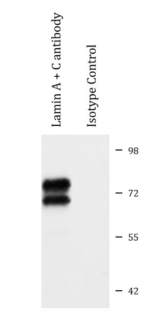
| Observed Size | 62 kDa, 74 kDa |
Properties
| Form | Liquid |
|---|---|
| Purification | Affinity purified. |
| Buffer | PBS (pH 7.3), 0.02% Sodium azide and 50% Glycerol. |
| Preservative | 0.02% Sodium azide |
| Stabilizer | 50% Glycerol |
| Storage Instruction | For continuous use, store undiluted antibody at 2-8°C for up to a week. For long-term storage, aliquot and store at -20°C. Storage in frost free freezers is not recommended. Avoid repeated freeze/thaw cycles. Suggest spin the vial prior to opening. The antibody solution should be gently mixed before use. |
| Note | For laboratory research only, not for drug, diagnostic or other use. |
Bioinformation
| Database Links | |
|---|---|
| Gene Symbol | LMNA |
| Gene Full Name | lamin A/C |
| Background | The nuclear lamina consists of a two-dimensional matrix of proteins located next to the inner nuclear membrane. The lamin family of proteins make up the matrix and are highly conserved in evolution. During mitosis, the lamina matrix is reversibly disassembled as the lamin proteins are phosphorylated. Lamin proteins are thought to be involved in nuclear stability, chromatin structure and gene expression. Vertebrate lamins consist of two types, A and B. Alternative splicing results in multiple transcript variants. Mutations in this gene lead to several diseases: Emery-Dreifuss muscular dystrophy, familial partial lipodystrophy, limb girdle muscular dystrophy, dilated cardiomyopathy, Charcot-Marie-Tooth disease, and Hutchinson-Gilford progeria syndrome. [provided by RefSeq, Apr 2012] |
| Function | Lamins are components of the nuclear lamina, a fibrous layer on the nucleoplasmic side of the inner nuclear membrane, which is thought to provide a framework for the nuclear envelope and may also interact with chromatin. Lamin A and C are present in equal amounts in the lamina of mammals. Plays an important role in nuclear assembly, chromatin organization, nuclear membrane and telomere dynamics. Required for normal development of peripheral nervous system and skeletal muscle and for muscle satellite cell proliferation. Required for osteoblastogenesis and bone formation. Also prevents fat infiltration of muscle and bone marrow, helping to maintain the volume and strength of skeletal muscle and bone. Prelamin-A/C can accelerate smooth muscle cell senescence. It acts to disrupt mitosis and induce DNA damage in vascular smooth muscle cells (VSMCs), leading to mitotic failure, genomic instability, and premature senescence. [UniProt] |
| Cellular Localization | Nucleus. Nucleus envelope. Nucleus lamina. Nucleus, nucleoplasm. Note=Farnesylation of prelamin-A/C facilitates nuclear envelope targeting and subsequent cleaveage by ZMPSTE24/FACE1 to remove the farnesyl group produces mature lamin-A/C, which can then be inserted into the nuclear lamina. EMD is required for proper localization of non-farnesylated prelamin-A/C. Isoform C: Nucleus speckle. [UniProt] |
| Calculated MW | Lamin A: 74 kDa Lamin C: 65 kDa |
| PTM | Increased phosphorylation of the lamins occurs before envelope disintegration and probably plays a role in regulating lamin associations. Proteolytic cleavage of the C-terminal of 18 residues of prelamin-A/C results in the production of lamin-A/C. The prelamin-A/C maturation pathway includes farnesylation of CAAX motif, ZMPSTE24/FACE1 mediated cleavage of the last three amino acids, methylation of the C-terminal cysteine and endoproteolytic removal of the last 15 C-terminal amino acids. Proteolytic cleavage requires prior farnesylation and methylation, and absence of these blocks cleavage. Sumoylation is necessary for the localization to the nuclear envelope. Farnesylation of prelamin-A/C facilitates nuclear envelope targeting. [UniProt] |
Images (4) Click the Picture to Zoom In
-
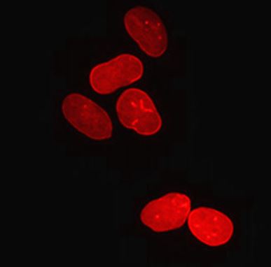
ARG42015 anti-Lamin A + C antibody ICC/IF image
Immunofluorescence: U2OS cells stained with ARG42015 anti-Lamin A + C antibody at 1:200 dilution.
-

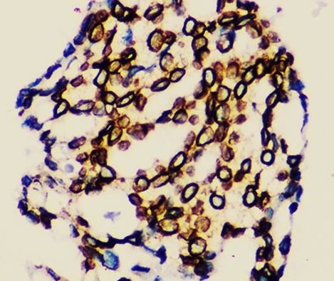
ARG42015 anti-Lamin A + C antibody IHC-P image
Immunohistochemistry: Paraffin-embedded Human breast cancer tissue stained with ARG42015 anti-Lamin A + C antibody at 1:200 dilution.
-
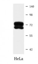
ARG42015 anti-Lamin A + C antibody WB image
Western blot: 25 µg of HeLa cell lysate stained with ARG42015 anti-Lamin A + C antibody at 1:10000 dilution.
-
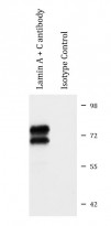
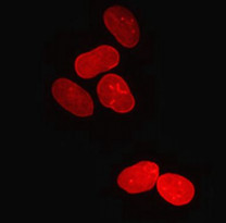
ARG42015 anti-Lamin A + C antibody IP image
Immunoprecipitation: 200 µg extracts of K562 cells were immunoprecipitated and stained with ARG42015 anti-Lamin A + C antibody at 1:20000 dilution.