ARG44389
anti-LEO1 antibody
anti-LEO1 antibody for ICC/IF,IHC-Formalin-fixed paraffin-embedded sections,Western blot and Human,Mouse,Rat
Overview
| Product Description | Rabbit Polyclonal antibody recognizes LEO1 |
|---|---|
| Tested Reactivity | Hu, Ms, Rat |
| Tested Application | ICC/IF, IHC-P, WB |
| Host | Rabbit |
| Clonality | Polyclonal |
| Isotype | IgG |
| Target Name | LEO1 |
| Antigen Species | Human |
| Immunogen | Recombinant fusion protein containing a sequence corresponding to amino acids 340-570 of human LEO1 |
| Conjugation | Un-conjugated |
| Alternate Names | LEO1; LEO1 Homolog, Paf1/RNA Polymerase II Complex Component; Replicative Senescence Down-Regulated Leo1-Like Protein; RNA Polymerase-Associated Protein LEO1; RDL; Leo1, Paf1/RNA Polymerase II Complex Component, Homolog (S. Cerevisiae); Leo1, Paf1/RNA Polymerase II Complex Component, Homolog; Leo1 Paf1/RNA Polymerase II Complex Component |
Application Instructions
| Application Suggestion |
|
||||||||
|---|---|---|---|---|---|---|---|---|---|
| Application Note | * The dilutions indicate recommended starting dilutions and the optimal dilutions or concentrations should be determined by the scientist. |
Properties
| Form | Liquid |
|---|---|
| Purification | Affinity purified. |
| Buffer | PBS (pH 7.3), 0.01% Thimerosal and 50% Glycerol. |
| Preservative | 0.01% Thimerosal |
| Stabilizer | 50% Glycerol |
| Storage Instruction | For continuous use, store undiluted antibody at 2-8°C for up to a week. For long-term storage, aliquot and store at -20°C. Storage in frost free freezers is not recommended. Avoid repeated freeze/thaw cycles. Suggest spin the vial prior to opening. The antibody solution should be gently mixed before use. |
| Note | For laboratory research only, not for drug, diagnostic or other use. |
Bioinformation
| Database Links | |
|---|---|
| Gene Symbol | LEO1 |
| Gene Full Name | LEO1 Homolog, Paf1/RNA Polymerase II Complex Component |
| Background | LEO1, parafibromin (CDC73; MIM 607393), CTR9 (MIM 609366), and PAF1 (MIM 610506) form the PAF protein complex that associates with the RNA polymerase II subunit POLR2A (MIM 180660) and with a histone methyltransferase complex. |
| Function | Component of the PAF1 complex (PAF1C) which has multiple functions during transcription by RNA polymerase II and is implicated in regulation of development and maintenance of embryonic stem cell pluripotency. PAF1C associates with RNA polymerase II through interaction with POLR2A CTD non-phosphorylated and 'Ser-2'- and 'Ser-5'-phosphorylated forms and is involved in transcriptional elongation, acting both independently and synergistically with TCEA1 and in cooperation with the DSIF complex and HTATSF1. PAF1C is required for transcription of Hox and Wnt target genes. PAF1C is involved in hematopoiesis and stimulates transcriptional activity of KMT2A/MLL1; it promotes leukemogenesis through association with KMT2A/MLL1-rearranged oncoproteins, such as KMT2A/MLL1-MLLT3/AF9 and KMT2A/MLL1-MLLT1/ENL. PAF1C is involved in histone modifications such as ubiquitination of histone H2B and methylation on histone H3 'Lys-4' (H3K4me3). PAF1C recruits the RNF20/40 E3 ubiquitin-protein ligase complex and the E2 enzyme UBE2A or UBE2B to chromatin which mediate monoubiquitination of 'Lys-120' of histone H2B (H2BK120ub1); UB2A/B-mediated H2B ubiquitination is proposed to be coupled to transcription. PAF1C is involved in mRNA 3' end formation probably through association with cleavage and poly(A) factors. In case of infection by influenza A strain H3N2, PAF1C associates with viral NS1 protein, thereby regulating gene transcription. Involved in polyadenylation of mRNA precursors. Connects PAF1C to Wnt signaling. |
| Cellular Localization | Nucleus |
| Calculated MW | 75 kDa |
| PTM | Acetylation, Phosphoprotein |
Images (7) Click the Picture to Zoom In
-

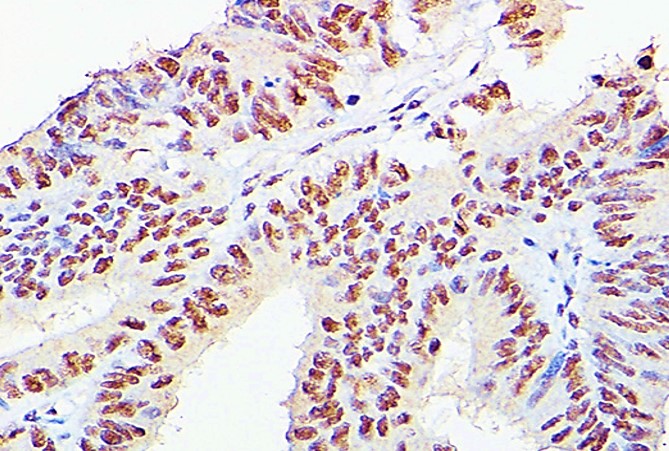
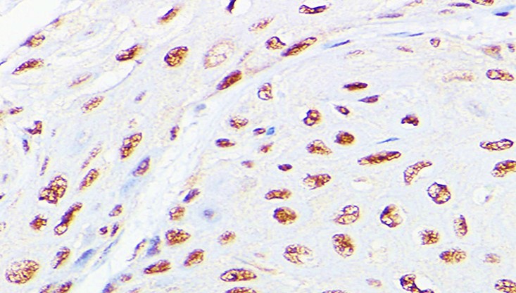
ARG44389 anti-LEO1 antibody IHC-P image
Immunohistochemistry: Human colon carcinoma stained with ARG44389 anti-LEO1 antibody at 1:100 dilution.
-
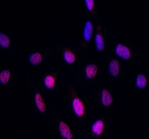
ARG44389 anti-LEO1 antibody ICC/IF image
Immunofluorescence: U2OS stained with ARG44389 anti-LEO1 antibody at 1:50 dilution.
-
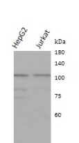
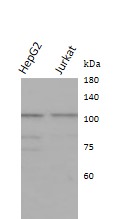
ARG44389 anti-LEO1 antibody WB image
Western blot: HepG2 and Jurkat stained with ARG44389 anti-LEO1 antibody at 1:1000 dilution.
-

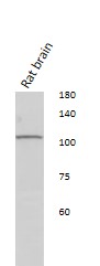
ARG44389 anti-LEO1 antibody WB image
Western blot: Rat brain stained with ARG44389 anti-LEO1 antibody at 1:1000 dilution.
-
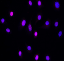
ARG44389 anti-LEO1 antibody ICC/IF image
Immunofluorescence: NIH/3T3 stained with ARG44389 anti-LEO1 antibody at 1:50 dilution.
-
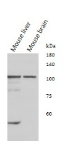
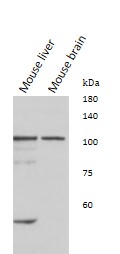
ARG44389 anti-LEO1 antibody WB image
Western blot: Mouse liver and Mouse brain stained with ARG44389 anti-LEO1 antibody at 1:1000 dilution.
-
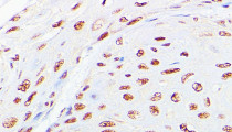
ARG44389 anti-LEO1 antibody IHC-P image
Immunohistochemistry: Human esophageal cancer stained with ARG44389 anti-LEO1 antibody at 1:100 dilution.